Image
Figure Caption
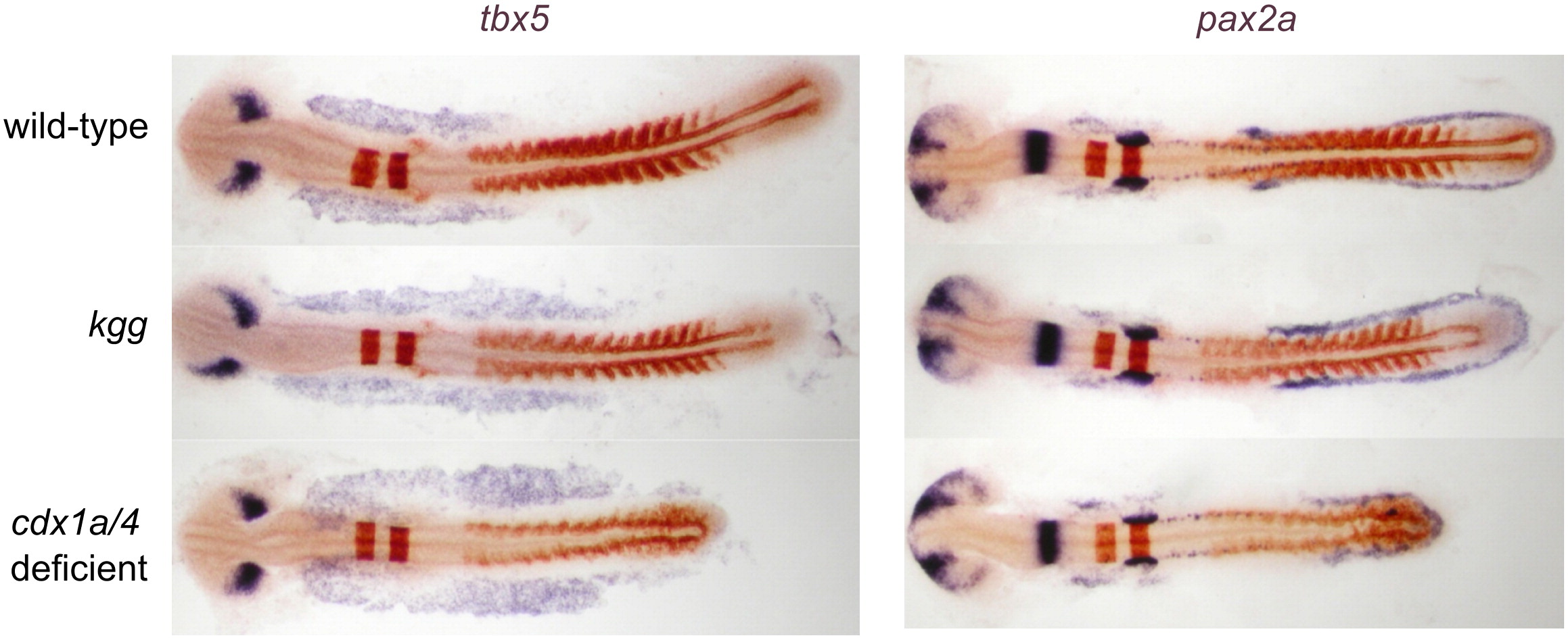
Fig. 5 Whole mount in situ hybridization analysis of tbx5a expression in wildtype, cdx4 and cdx1a/4-deficient embryos shows expansion of cardiogenic anterior-lateral plate mesoderm. The intermediate mesoderm field, marked by pax2a expression, shows a posterior shift and reduction in cell number, as reported previously. The effect was more pronounced in cdx1/4 double deficient embryos than in cdx4-/- embryos. Shown are zebrafish embryos at 15-somite stage. Purple staining was used for tbx5 and pax2a and red staining for krox20 and myoD as landmarks of other tissues.
Figure Data
Acknowledgments
This image is the copyrighted work of the attributed author or publisher, and
ZFIN has permission only to display this image to its users.
Additional permissions should be obtained from the applicable author or publisher of the image.
Reprinted from Developmental Biology, 354(1), Lengerke, C., Wingert, R., Beeretz, M., Grauer, M., Schmidt, A.G., Konantz, M., Daley, G.Q., and Davidson, A.J., Interactions between Cdx genes and retinoic acid modulate early cardiogenesis, 134-142, Copyright (2011) with permission from Elsevier. Full text @ Dev. Biol.

