
|
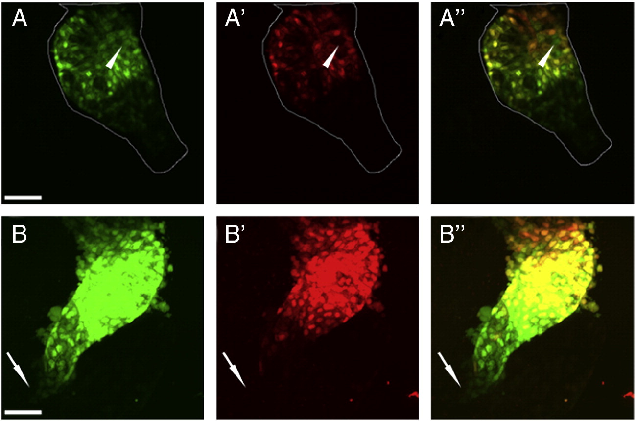
Fig. S2 Late myocardial addition occurred rarely in the distal ventricle; newest myl7-positive cells were found distal to the arterial pole. (A–A′′) Single z-stack of 48 hpf heart exposed to UV at 30 hpf, with outline marking the ventricle. White arrowhead points to green fluorescing cells (A) that did not fluoresce red (A′). Overlay (A′′) showed these cells to be separate from green-only cell in arterial pole. (B–B′′) Over-exposure of 48 hpf heart exposed to UV at 30 hpf. White arrow points to faint green-only cells visible under high exposure (B), that did not fluoresce red (B′ and B′′ for overlay). Green channel (A and B), red channel (A′ and B′), and overlay (A′′ and B′′). Scale bar represents 50 μm.
Reprinted from Developmental Biology, 354(1), Lazic, S., and Scott, I.C., Mef2cb regulates late myocardial cell addition from a second heart field-like population of progenitors in zebrafish, 123-133, Copyright (2011) with permission from Elsevier. Full text @ Dev. Biol.