
Image
|
Figure Caption
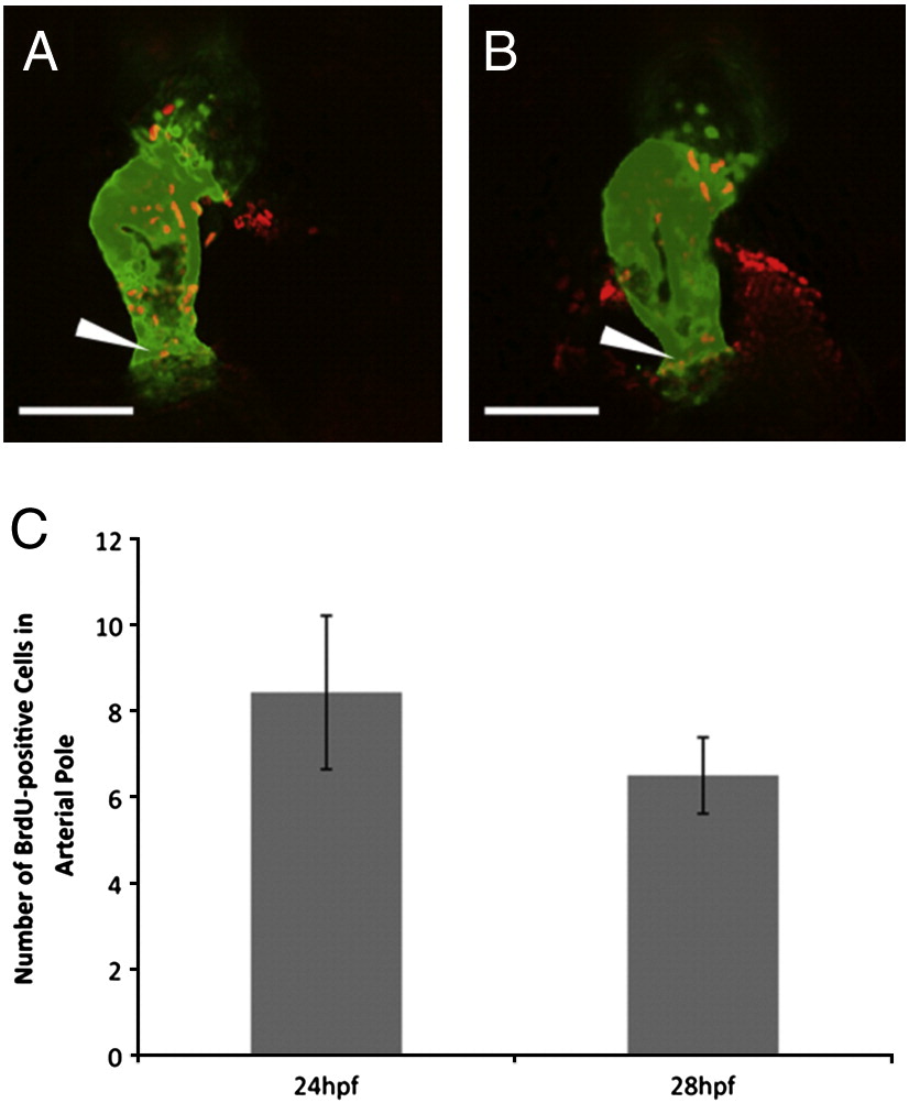
Fig. 7 Cell proliferation in the arterial pole. myl7:EGFP embryos were pulsed with BrdU for 1 h at 24 hpf (A) and 28 hpf (B) and cell proliferation was assessed at 48 hpf. The myocardium is visualized in the green channel and BrdU labeled cells are visualized in the red channel. Proliferating cells in the arterial pole of the heart (arrowheads) were quantified (C). Scale bar represents 70 μm.
Acknowledgments
This image is the copyrighted work of the attributed author or publisher, and
ZFIN has permission only to display this image to its users.
Additional permissions should be obtained from the applicable author or publisher of the image.
Reprinted from Developmental Biology, 354(1), Lazic, S., and Scott, I.C., Mef2cb regulates late myocardial cell addition from a second heart field-like population of progenitors in zebrafish, 123-133, Copyright (2011) with permission from Elsevier. Full text @ Dev. Biol.