
|
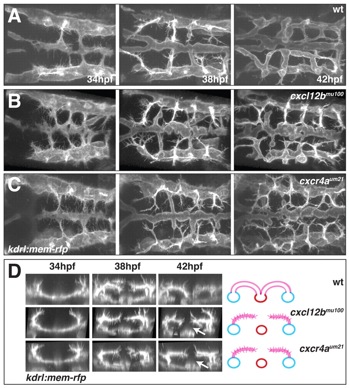
Fig. 5
Time-lapse imaging of hindbrain vascular development in wt, cxcl12bmu100 and cxcr4aum21 mutant embryos. (A-C) Still images at 34, 38 and 42 hpf from confocal time-lapse movies of hindbrain vascular development in live wt (A), cxcl12bmu100 (B) and cxcr4aum21 (C) mutant kdrl:mem-rfp transgenic embryos (see Movies 7-9 in the supplementary material). Dorsal views, anterior to the left. (D) Transverse sections based on 3D reconstruction of the movies illustrated in A-C, highlighting the absence of ventral migration of CtA sprouts in cxcl12bmu100 and cxcr4aum21 mutant embryos (arrows). The schematic interpretations are based on 42 hpf still images as in Fig. 1I.