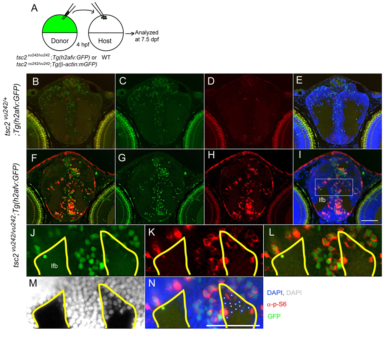
Fig. 6 Cell-autonomous activation of TORC1 in tsc2vu242/vu242 mutant cells and non-cell-autonomous disruption of white matter. (A) Schematic of mosaic analyses: cells from wild-type tsc2;Tg(h2afv:GFP) or tsc2vu242/vu242;Tg(h2afv:GFP) donors were transplanted into wild-type host blastulae at 4 hpf, and host embryos were analyzed at 7.5 dpf. (B–L) Coronal brain sections from wild-type embryos receiving either Tg(h2afv:GFP) wild-type donor cells (B–D) or tsc2vu242/vu242; Tg(h2afv:GFP) mutant donor cells (E–L). (C,G) Green (GFP); (D,H) red (phospho-S6); (E,I) green (GFP), red (phospho-S6), blue (DAPI) merged images. (B) Merged image of C and D; (F) merged image of G and H to delineate transplanted cells and those with increased mTORC1 signaling. (J–N) Magnified views of rectangle in I. Asterisks point to wild-type host cells (GFP negative and phospho-S6 negative), which seem to be ectopically positioned within the white matter (M,N). (J–N) The area outlined in yellow marks the normal gray-white matter limits. lfb, lateral forebrain bundle. Scale bars: 50 μm.
Image
Figure Caption
Acknowledgments
This image is the copyrighted work of the attributed author or publisher, and
ZFIN has permission only to display this image to its users.
Additional permissions should be obtained from the applicable author or publisher of the image.
Full text @ Dis. Model. Mech.

