Image
Figure Caption
Fig. S2
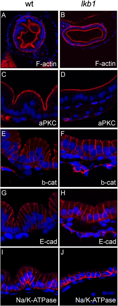
There were no polarity defects in the intestine of lkb1-mutant larvae. Immunofluorescent analysis of transverse sections of the intestinal bulb in WT larvae (A, C, E, G, I, K) and ikb1-mutant larvae (B, D, F, H, J, L) at 7 dpf. Apical localization and levels of F-actin (A and B) and atypical PKC (aPKC) (C and D) are normal in lkb1-mutant larvae. Basolateral localization of β-catenin (E and F), E-cadherin (G and H), and Na/K ATPase (I and J), is not affected in lkb1-mutant larvae.
Acknowledgments
This image is the copyrighted work of the attributed author or publisher, and
ZFIN has permission only to display this image to its users.
Additional permissions should be obtained from the applicable author or publisher of the image.
Full text @ Proc. Natl. Acad. Sci. USA

