
|
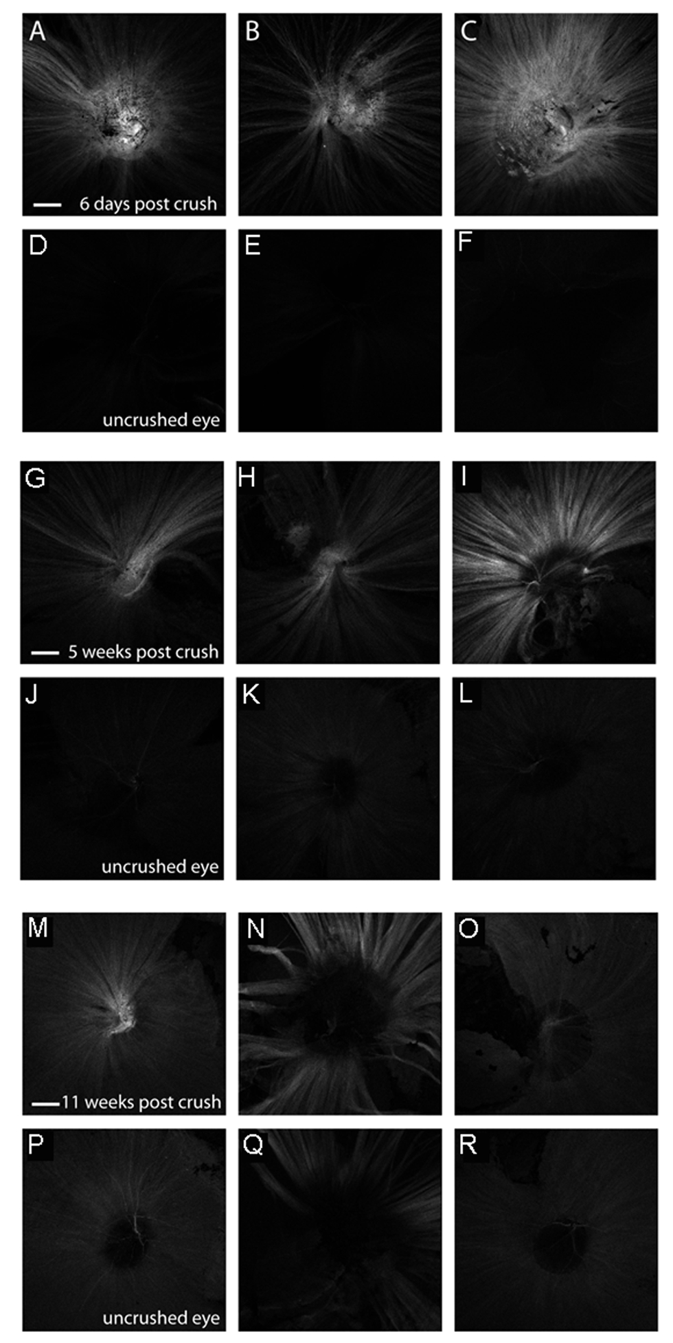
Fig. s4
Expression of the gap43:GFP transgene following optic nerve crush in adult (6 month old) wild-type fish. A-F Expression of the gap43:GFP transgene in experimental eyes 6 days after optic nerve crush (A-D) or in the contralateral uncrushed control eye (E-F). G-L Expression of the gap43:GFP transgene in experimental eyes 5 weeks after optic nerve crush (I-L) or in the contralateral uncrushed control eyes (M-P). M-R Expression of the gap43:GFP transgene in experimental eyes 11 weeks after optic nerve crush (Q-T) or in the contralateral uncrushed control eyes (E-F). Shown are 3 representative samples for each condition. Images capture a single plane of the nerve fiber layer using equal gain settings on a confocal microscope. Scale bar = 200μm.