
|
Fig. 2
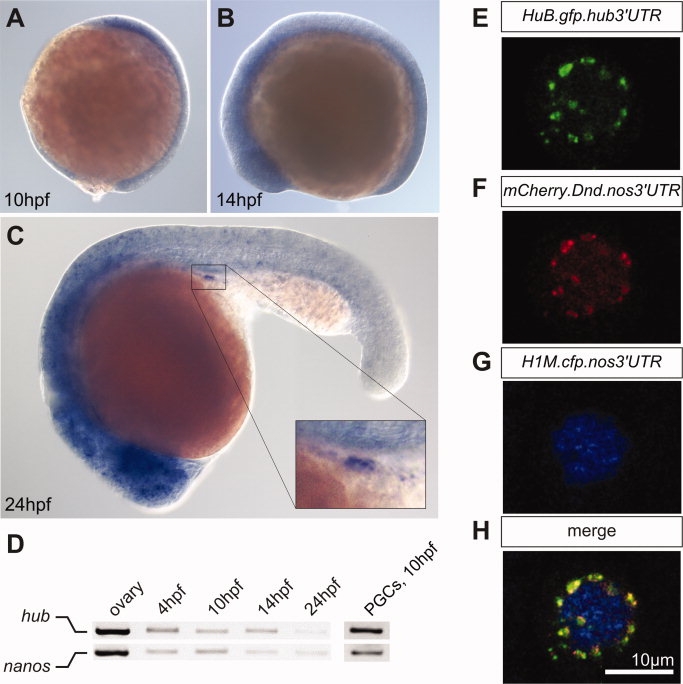
Figure 2. hub mRNA is expressed in the head and in PGCs of zebrafish embryos and HuB protein is localized to perinuclear granules in the germ cells. A?C: Whole-mount in situ hybridization of wild type zebrafish embryos using an antisense probe directed against zebrafish hub RNA at the indicated developmental stages. hub mRNA is ubiquitously expressed in 10-hpf (A) and in 14-hpf embryos (B). In 24-hpf embryos, hub RNA is expressed in the head and in PGCs (inset) (C). D: The expression of nanos and hub mRNA determined by RT-PCR. Ovary cDNA, cDNA of embryos of the indicated developmental stages, and cDNA of germ cells isolated from 10hpf embryos were used as templates. E?H: Confocal microscopy images of a PGC in a 6hpf embryo showing the sub-cellular localization of the HuB-GFP fusion protein. The HuB fusion protein is localized to granular structures around the nucleus (E), which are co-labeled with the mCherry-Dnd fusion protein (F). The nucleus is marked by the CFP-histone protein (G) and the merged image is shown in H.