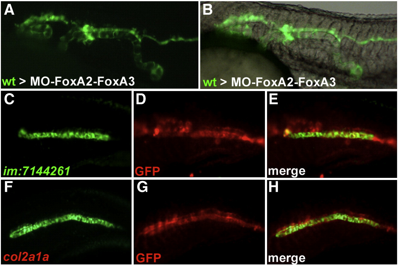
Fig. 6 Wild-type cells transplanted into FoxA2?FoxA3 morphant embryos form axial structures in a cell autonomous manner. (A and B) Trunk region of FoxA2?FoxA3 morphants at 24 hpf after transplantation of wild-type cells labeled with GFP. Wild-type cells form stretches of axial structures composed exclusively of transplanted cells. (C?H) Identity of the transplanted cells analyzed by labeling of GFP (D and G) with the notochord marker im:7144261 (C) or the floor plate and hypochord marker col2a1a (F). All cells expressing col2a1a and im:7144261 are also labeled for GFP (E and H). Lateral views, anterior to the left.
Reprinted from Developmental Biology, 350(2), Dal-Pra, S., Thisse, C., and Thisse, B., FoxA transcription factors are essential for the development of dorsal axial structures, 484-495, Copyright (2011) with permission from Elsevier. Full text @ Dev. Biol.

