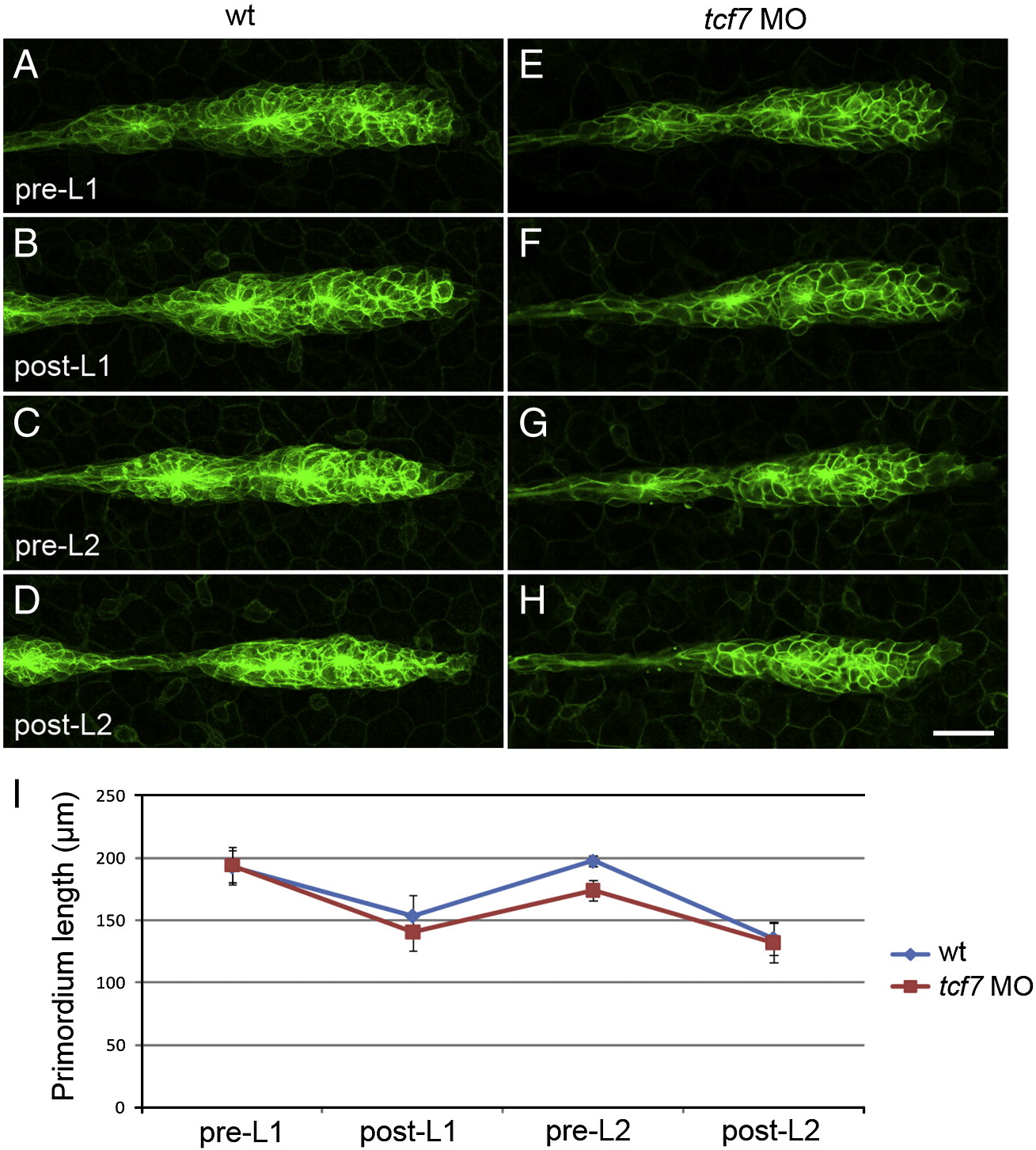
Fig. 4 Primordium lengthening precedes proneuromast deposition. Still images of timelapse movies of proneuromast deposition in control and tcf7 MO injected Tg(claudinb:GFP) transgenic embryos (A?H). (A?D) Wt primordia in different phases of the deposition cycle. (A) Wt primordium immediately prior to deposition of the first primary proneuromast (L1). (B) The same primordium immediately following deposition of L1. (C) Wt primordium preceding deposition of the second proneuromast (L2). (D) Wt primordium following deposition of L2. (E,F) tcf7 morphant primordia before deposition and following deposition of L1. (G,H) The same primordium immediately before and following deposition of L2. (I) Quantification of primordium lengths in wt embryos (blue, n = 4) and tcf7 morphants (red, n = 7). Wt and tcf7 morphant primordia achieve similar lengths preceding deposition of L1 (A,E,I). Wt and tcf7 morphant primordia shrink to similar sizes following deposition of L1 (B,F,I). tcf7 morphant primordia may be slightly shorter than wt primordia preceding L2 deposition (C,G,I). However, following deposition of L2, wt and tcf7 morphant primordia again possess the same length (D,H,I).
Reprinted from Developmental Biology, 349(2), Aman, A., Nguyen, M., and Piotrowski, T., Wnt/?-catenin dependent cell proliferation underlies segmented lateral line morphogenesis, 470-482, Copyright (2011) with permission from Elsevier. Full text @ Dev. Biol.

