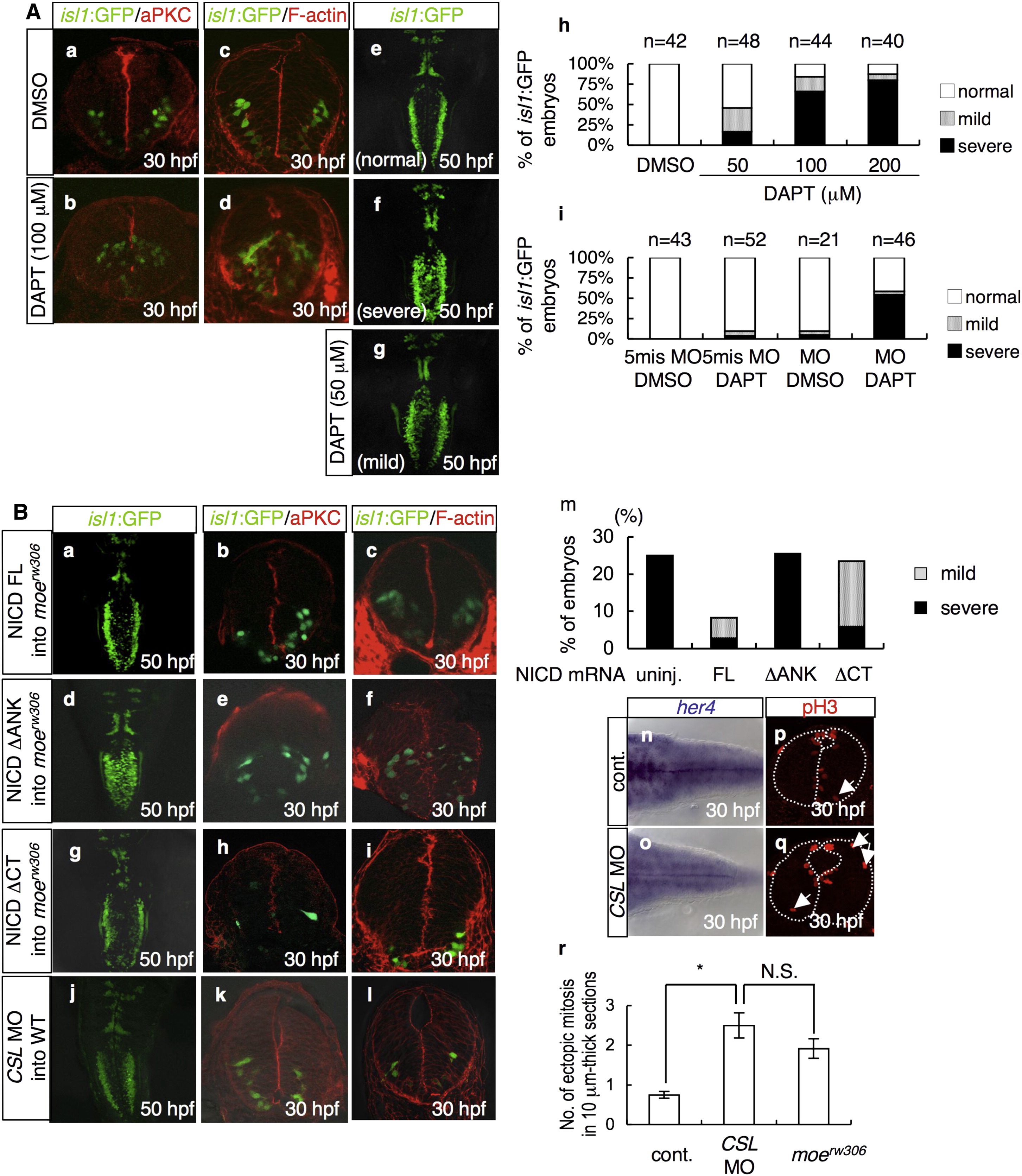
Fig. 6 Notch Functions Downstream of Moe to Maintain Neuroepithelial Apicobasal Polarity
(A) Visualization of aPKC (a and b) and F-actin (c and d) at 30 hpf, and dorsal views (e, f, and g) at 50 hpf of the embryos treated with DMSO (a, c, and e; 1.0%) or DAPT (b, d, and f; 100 μM: g; 50 μM) from 6 hpf to the indicated developmental stages. (h and i) Percentages of isl1:GFP embryos with bilaterally segregated vagus motor nuclei (normal, open bars, e), partially fused vagus motor nuclei (mild, gray bars, g), and completely fused vagus motor nuclei (severe, black bars, f) at 48 hpf. (h) The isl1:GFP embryos were treated with DMSO (2.0%) or DAPT at the indicated concentrations from 6 hpf to 48 hpf. (i) The isl1:GFP embryos were injected with the 5-mis-pair control MO (5-mis MO; 0.20 mg/ml) or moe MO (MO; 0.20 mg/ml) and then treated with DMSO (0.20%) or DAPT (20 μM) from 6 hpf to 48 hpf.
(B) (a?l) The moerw306 mutant embryos (a?i) were injected with the NICD FL (a?c; 60 μg/ml), NICD ΔANK (d?f; 60 μg/ml), and NICD ΔCT (g?i; 20 μg/ml) mRNA species. The WT embryos were injected with CSL MO (j?l; 0.20 mg/ml). These embryos were examined for the formation of the vagus motor nuclei (a, d, g, and j; dorsal views, 50 hpf), neuroepithelial polarity (b, e, h, and k; aPKC, cross-sectional views, 30 hpf), and intercellular junctions (c, f, i, and l; F-actin, cross-sectional views, 30 hpf). (m) Percentages of embryos with partially fused vagus motor nuclei (gray bars) and completely fused vagus motor nuclei (black bars) at 48 hpf. The embryos obtained from moerw306 heterozygous mutant fish pairs underwent no injection (uninj.) or injection with the NICD FL (60 μg/ml), NICD ΔANK (60 μg/ml), and NICD ΔCT (20 μg/ml) mRNA species. (n?r) In situ hybridization of her4 (n and o) and immunohistochemistry of pH3 (p and q) were assessed in the WT embryos injected with control MO (n and p; 0.20 mg/ml) and CSL MO (o and q; 0.20 mg/ml). The number of ectopically dividing cells was assessed in 10 μm thick sections (r). The data shown in (r) are mean ± SEM; *p < 0.05.

