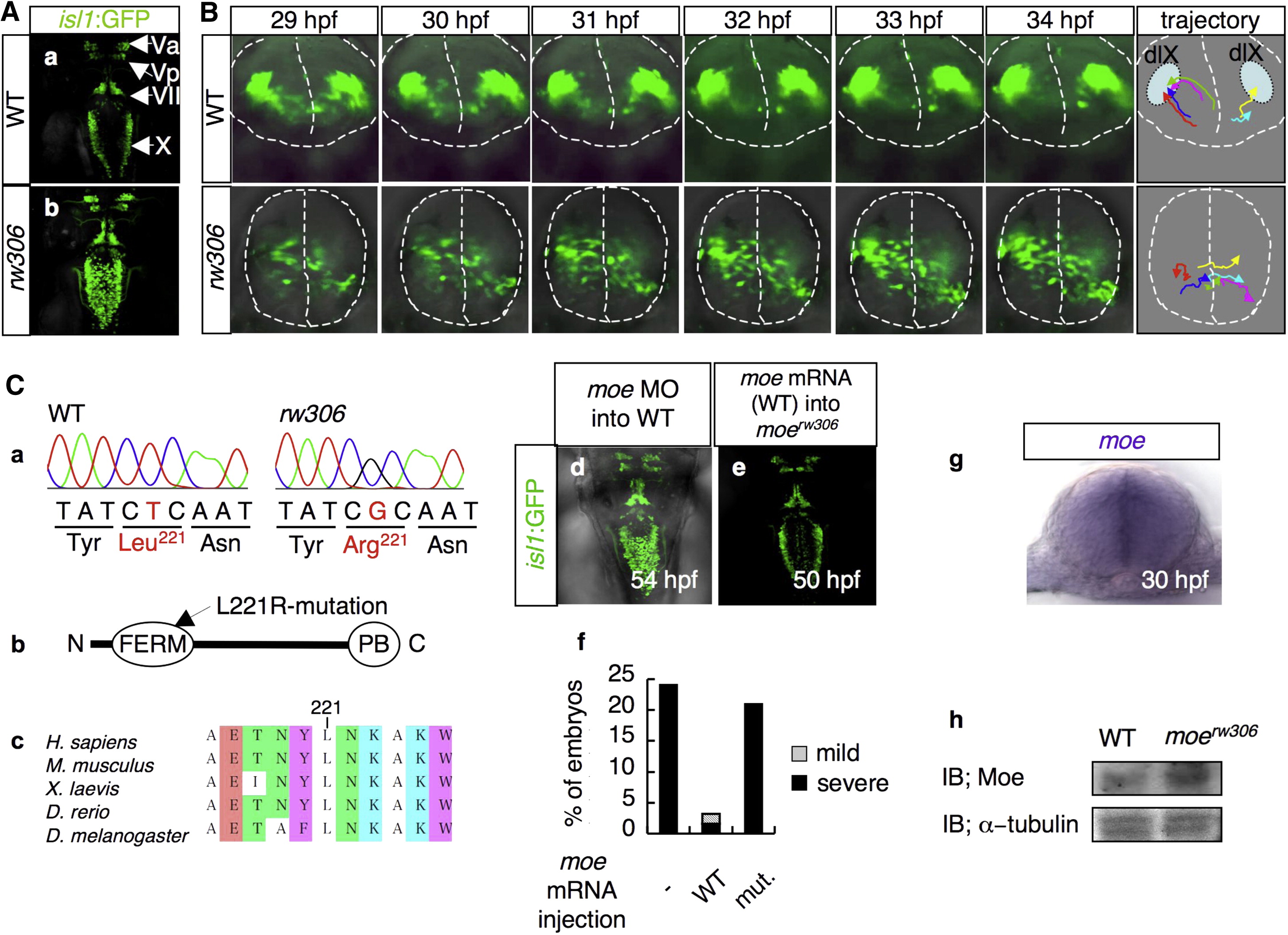
Fig. 1 The moerw306 Mutation Affects the Directionality of the Migration of the Vagus Motor Neuron Precursors
(A) Fluorescence images of the hindbrains of isl1:GFP transgenic zebrafish at 50 hpf. (a) WT and (b) rw306 mutant. Dorsal view (rostral toward the top) is shown. The following abbreviations are used: Va, anterior trigeminal motor nuclei; Vp, posterior trigeminal motor nuclei; VII, facial motor nuclei; X, vagus motor nuclei.
(B) Migration patterns of the vagus motor neuron precursors in the WT (upper) and rw306 mutant (lower) from 29 hpf to 34 hpf. The dotted lines indicate the outlines of the neural tubes and the midlines. The colored arrows in the trajectory panel indicate the trajectories of six arbitrarily chosen vagus motor neuron precursors. dlX is an abbreviation for dorsolateral motor nucleus of the vagus.
(C) (a) Sequences of the mutation sites in the WT allele and the rw306 allele. (b) Primary structure of Moe. The arrow indicates the position of the L221R mutation in Moe. The following abbreviations are used: N, N terminus; C, C terminus; PB, PDZ-binding domain. (c) Alignment of the Moe amino acid sequences from various species. The amino acids are colored according to type: blue, polar positive (K); green, polar neutral (T, N); white, nonpolar aliphatic (A, L, I); purple, nonpolar aromatic (Y, W); brown, other (E). (d and e) Dorsal views of the WT embryo injected with moe MO (d; 2.0 mg/ml) at 54 hpf and the moerw306 mutant embryo injected with WT moe mRNA (e; 100 μg/ml) at 50 hpf (rostral to the top). (f) Percentages of embryos with partially fused vagus motor nuclei (gray bars) and completely fused vagus motor nuclei (black bars) at 48 hpf. The embryos obtained from moerw306 heterozygous mutant fish pairs underwent no injection (-), injection with the WT moe mRNA (WT; 100 μg/ml), or injection with the L221R-type moe mRNA (mut; 100 μg/ml). (g) Expression pattern of moe mRNA in the hindbrain at 30 hpf (cross-sectional view, dorsal to the top). (h) Western blotting for detection of the Moe protein in the WT and moerw306 mutant embryos; immunoreactivity for α-tubulin is shown as a loading control. IB is an abbreviation for immunoblot.

