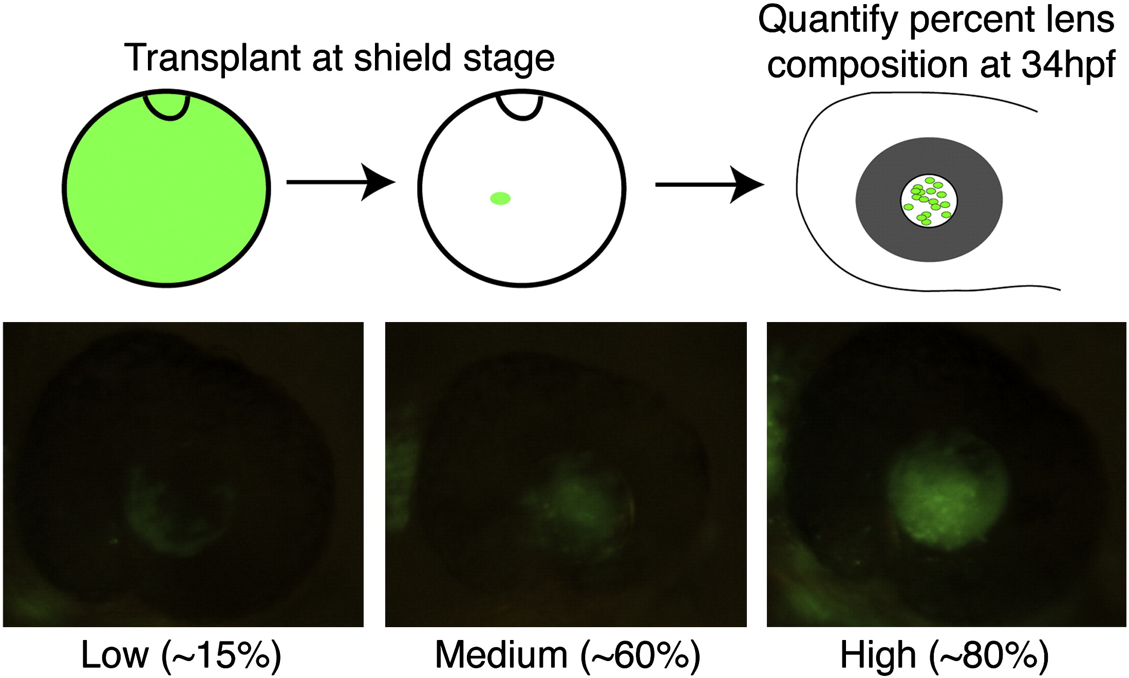
Fig. S6 Shield-stage lens mosaics. Cartoon schematic of shield-stage transplants. Donor embryos are injected with Alexa Fluor 488 dextran (10 kDa) and grown to the shield stage. Animal pole view of shield-stage host embryos (shield is located at 12 o′clock in the cartoon), indicating the approximate location of the lens-fated region of the embryo into which donor cells were transplanted. Embryos were grown to 34 hpf and screened for percent lens composition and classed into ?Low? (< 30% donor cells), ?Medium? (30?70% donor cells) or ?High? (> 70% donor cells) groups. Embryos with any retinal contamination were discarded. The genotype of donor and host embryos was determined at 5 dpf based on phenotype, and only those involving severe mutants were used for subsequent analyses.
Reprinted from Developmental Biology, 350(1), Tittle, R.K., Sze, R., Ng, A., Nuckels, R.J., Swartz, M.E., Anderson, R.M., Bosch, J., Stainier, D.Y., Eberhart, J.K., and Gross, J.M., Uhrf1 and Dnmt1 are required for development and maintenance of the zebrafish lens, 50-63, Copyright (2011) with permission from Elsevier. Full text @ Dev. Biol.

