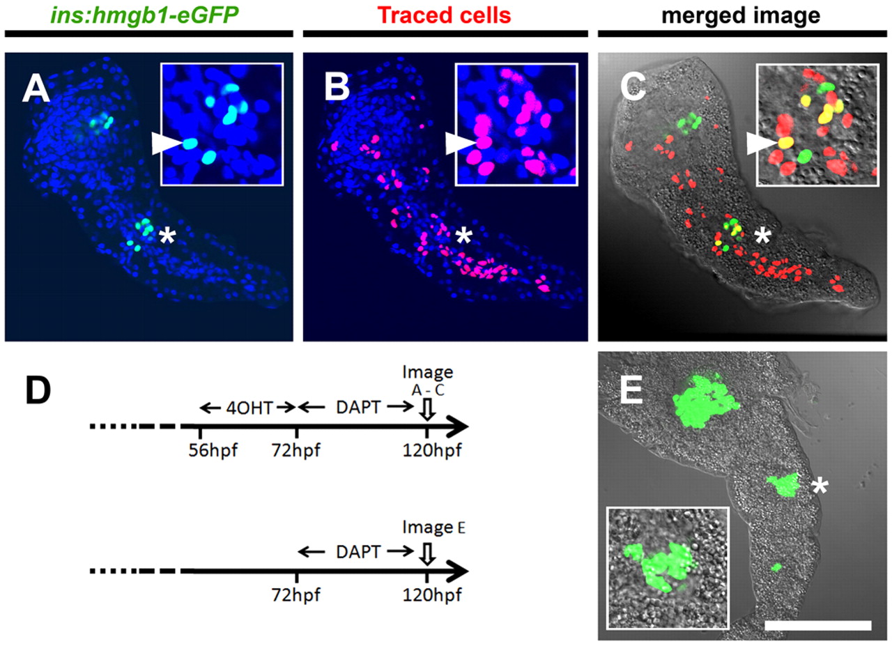
Fig. 5 Pancreatic Notch-responsive progenitors form precocious secondary islets. (A-C,E) Confocal images of single optic sections of micro-dissected pancreata from GIFM; ins:hmgb1-eGFP larvae at 5 dpf. PNCs were labeled by addition of 4OHT from 56 to 72 hpf. All larvae were incubated in DAPT from 72 to 120 hpf, which induces precocious secondary islets. (A) Nuclear GFP expression shows position of β-cells. (B) Nuclear mCherry labels the PNCs and maps their subsequent developmental fate. All nuclear counterstained with DAPI (blue). (C) In red/green images merged with bright-field images, co-labeled cells appear yellow. (A-C) Position of the secondary islet is marked (*) and magnified in the inset panels, where an example of a lineage trace β-cell is marked with a white arrowhead. (D) Schematic of 4OHT-dependent fate mapping and negative control (no 4OHT) in GIFM fish with secondary islet induction. (E) Without 4OHT treatment, no fate mapping is observed. Scale bar: 100 μm
Image
Figure Caption
Acknowledgments
This image is the copyrighted work of the attributed author or publisher, and
ZFIN has permission only to display this image to its users.
Additional permissions should be obtained from the applicable author or publisher of the image.
Full text @ Development

