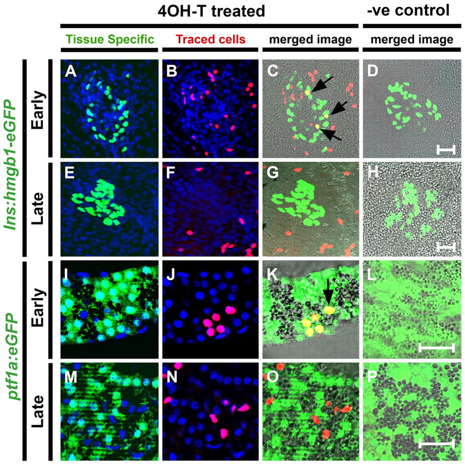
Fig. 3 Early Notch-responsive progenitors contribute to principal islet β-cells and exocrine cells. (A-P) Confocal images (single optic section) of microdissected pancreata from GIFM larvae at 5 dpf. Fish are also transgenic for either (A-H) ins:hmgb1-eGFP (marking β-cells with nuclear GFP) or (I-P) ptf1a:eGFP (marking acinar cells with GFP). As indicated, larvae were treated either with 4OHT (4OHT treated) or with vehicle alone as a negative control (–ve control; D,H,L,P). 4OHT or vehicle was added at two different stages of development: 12-36 hpf, which is a developmental stage covering the appearance of the pancreas (Early); or 56-80 hpf, a stage of pancreas maturation (Late). (A,E,I,M) Tissue-specific markers in GFP and nuclei in DAPI (blue). (B,F,J,N) Lineage-traced cells with red labeled nuclei. (C,G,K,O) Red and green images merged with bright field. Cells positive for both tissue-specific transgene and lineage labeling appear yellow; examples are indicated with black arrows. Treatment with 4OHT leads to labeling of β-cells of the principal islet and cells of the exocrine pancreas when added at 12 hpf (C,K) but rarely if added later at 56 hpf (G,O). Scale bars: 20 μm in D,H,L,P.
Image
Figure Caption
Acknowledgments
This image is the copyrighted work of the attributed author or publisher, and
ZFIN has permission only to display this image to its users.
Additional permissions should be obtained from the applicable author or publisher of the image.
Full text @ Development

