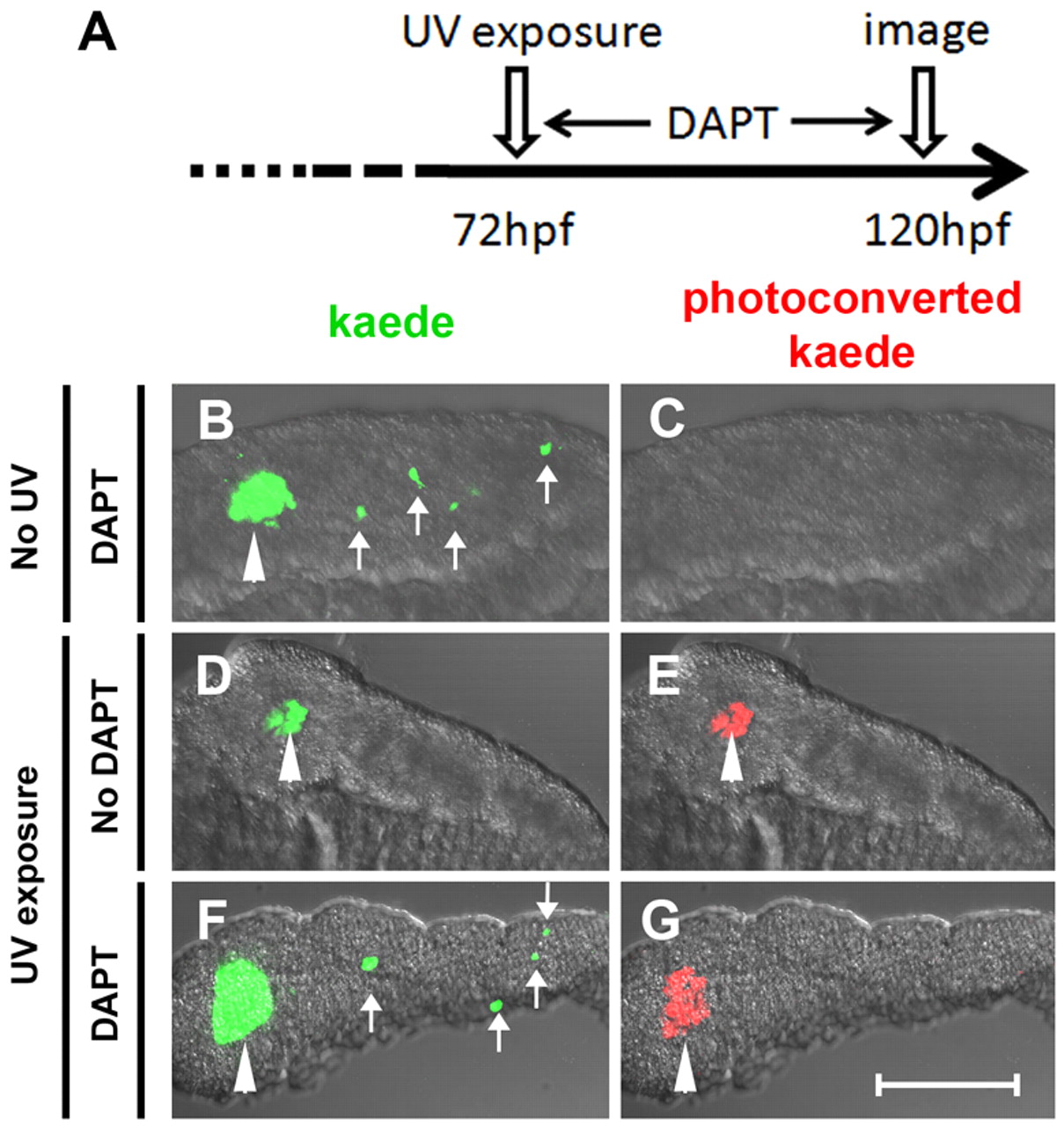
Fig. 1 β-Cells of the secondary islets do not originate from principal islet β-cells. (A) Schematic of experimental timeline. The green fluorescent kaede protein in β-cells of ins:kaede fish was photoconverted to red fluorescent protein by UV exposure at 72 hpf. (B-G) Rendered confocal images of microdissected pancreata from ins:kaede fish at 120 hpf. (B,F) DAPT treatment from 72-120 hpf induces secondary islets (white arrows), including differentiated β-cells that are marked by kaede protein. (E,G) Photoconversion at 72 hpf labeled all β-cells present at that time point with red fluorescence. By 120 hpf, photoconverted kaede is still apparent in principal islet cells (white arrowheads) but is not detected in DAPT-induced secondary islets (G). Scale bar: 100 μm.
Image
Figure Caption
Acknowledgments
This image is the copyrighted work of the attributed author or publisher, and
ZFIN has permission only to display this image to its users.
Additional permissions should be obtained from the applicable author or publisher of the image.
Full text @ Development

