
|
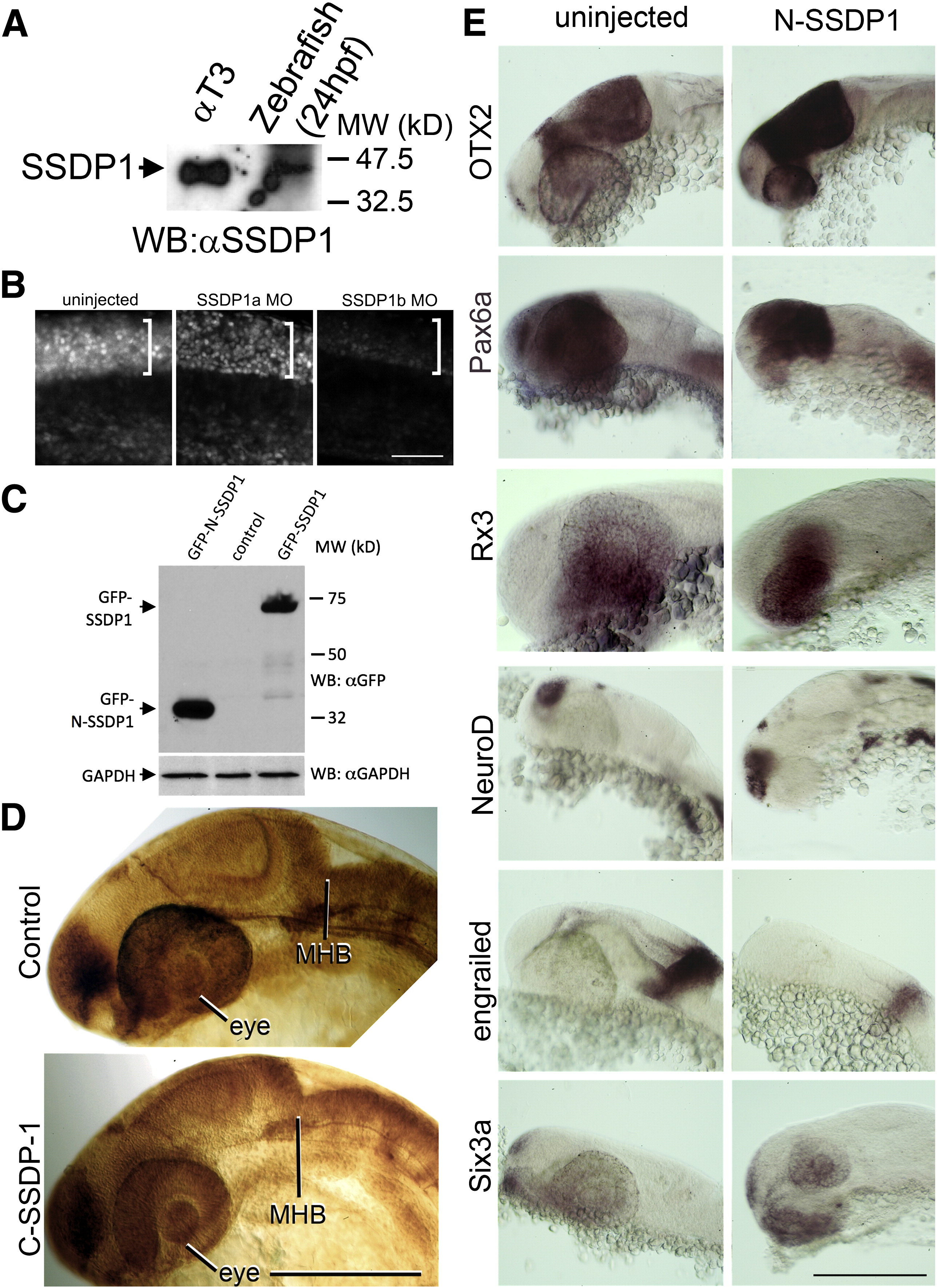
Fig. S2 A: Western blot analysis indicates that the SSDP1 antibody detects a band of the same molecular weight in protein extracts of αT3 cells and zebrafish embryos, suggesting specific reactivity of the antibody in zebrafish embryos. B: Immunodetectability of SSDP1 is slightly reduced by SSDP1a and strongly reduced by SSDP1b morpholino injection, indicating that the morpholino predominantly recognizes SSDP1b. Lateral trunk views of whole-mounted embryos are shown. The strongly immunoreactive spinal cord level is indicated by brackets. C: Western blot analysis indicates that GFP-tagged N-SSDP1 and full length SSDP1 can be expressed in αT3 cells. D: Lateral views of anti-tubulin labeled embryos at 24 hpf are shown. Over-expression of C-SSDP1, which lacks the CLIM-interacting domain, does not induce phenotypes in eye or MHB. E: Lateral views of the heads of 24 hpf embryos after in situ hybridization are shown. The indicated markers retain their general expression patterns when N-SSDP1 is over-expressed. Scale bar in B = 50 μm; bars in D,E = 200 μm.
Reprinted from Developmental Biology, 349(2), Zhong, Z., Ma, H., Taniguchi-Ishigaki, N., Nagarajan, L., Becker, C.G., Bach, I., and Becker, T., SSDP cofactors regulate neural patterning and differentiation of specific axonal projections, 213-224, Copyright (2011) with permission from Elsevier. Full text @ Dev. Biol.