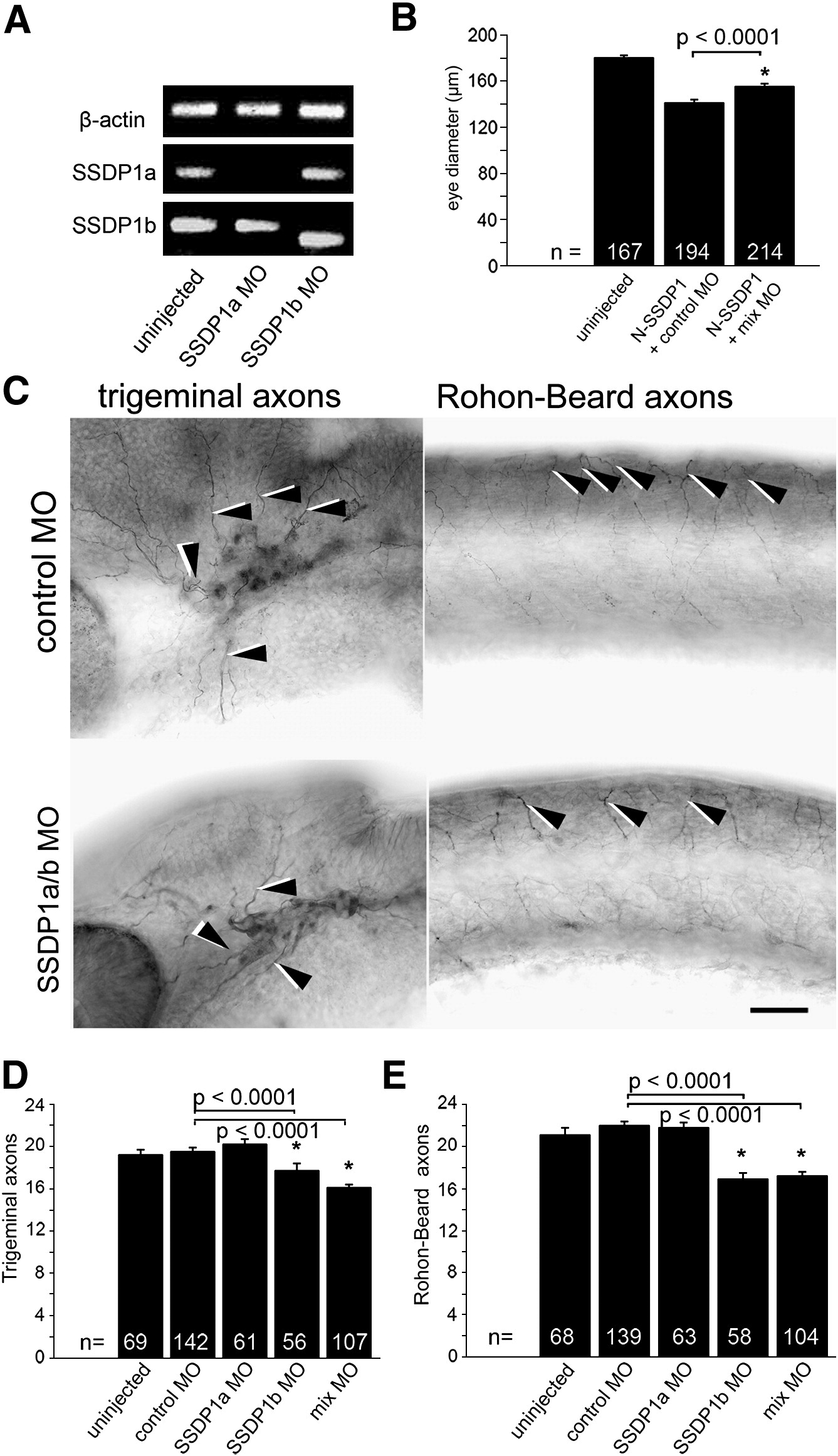
Fig. 6 Knock down of SSDP1b, but not SSDP1a partially inhibits growth of peripheral axons of trigeminal and Rohon?Beard neurons. A: Morpholino knock down reduces levels of correctly spliced SSDP1a and SSDP1b to levels that are undetectable by PCR. An ectopic band is amplified after SSDP1b knock down, which contains premature stop codons and, therefore, cannot lead to functional protein. Beta-actin was used to equalize cDNA concentrations. B: Morpholinos to SSDP1 partially rescue reduced eye size induced by N-SSDP1 over-expression, suggesting specificity of the morpholinos. C: Lateral views of anti-tubulin immuno-labeled whole-mounted 24 hpf embryos injected with morpholinos against SSDP1a/b are shown. Arrowheads point to trigeminal and Rohon?Beard axons, respectively. Scale bar = 50 μm. D, E: Quantifications indicate significant loss of peripheral axons of trigeminal and Rohon?Beard neurons after morpholino knock down of SSDP1b.
Reprinted from Developmental Biology, 349(2), Zhong, Z., Ma, H., Taniguchi-Ishigaki, N., Nagarajan, L., Becker, C.G., Bach, I., and Becker, T., SSDP cofactors regulate neural patterning and differentiation of specific axonal projections, 213-224, Copyright (2011) with permission from Elsevier. Full text @ Dev. Biol.

