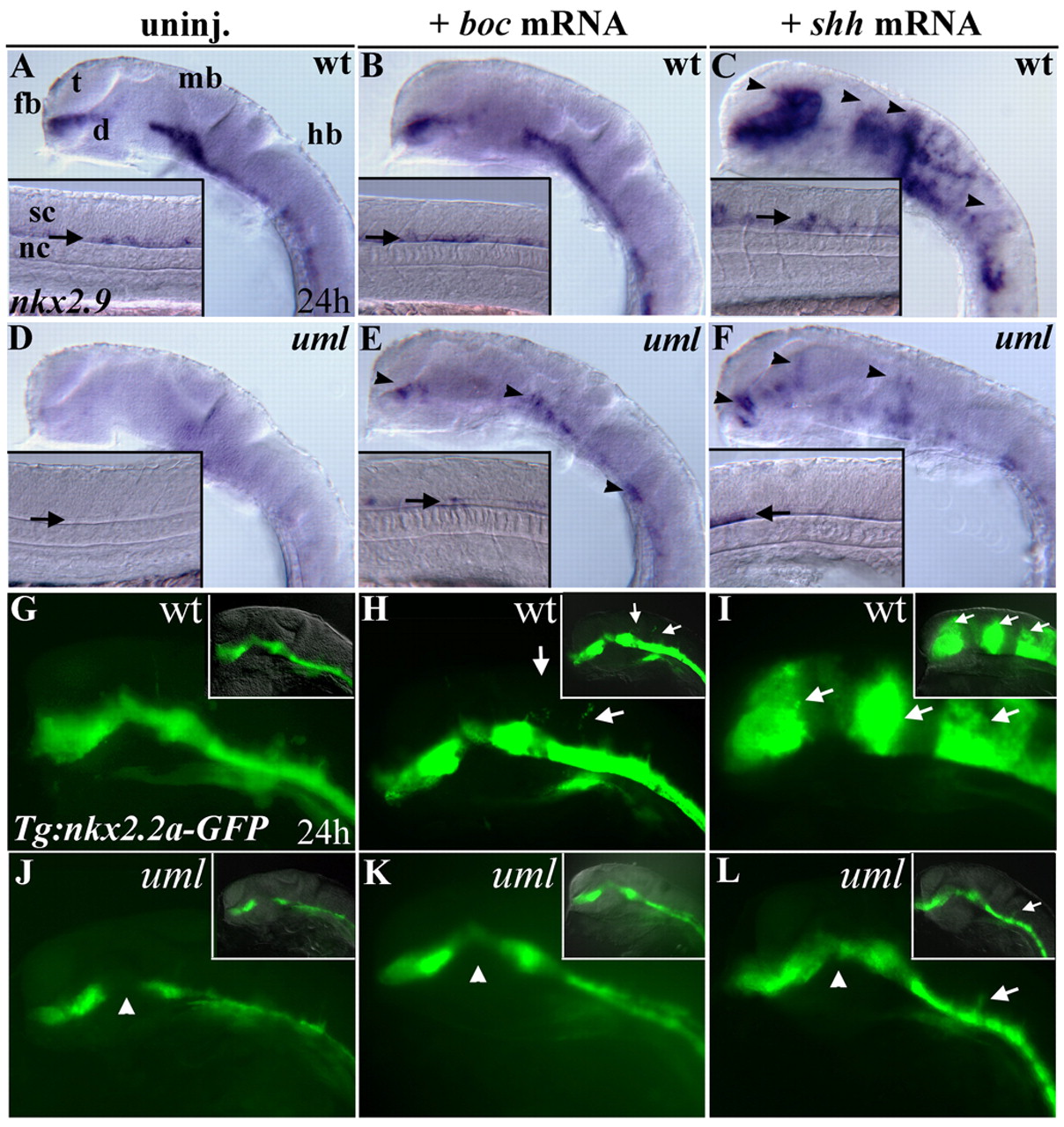
Fig. 5 boc mRNA injections rescue Hh signaling defects in uml mutants and weakly activate Hh signaling ectopically. (A) Wild-type nkx2.9 expression in the brain and lateral floor plate (inset, arrow). (B) Ectopic nkx2.9 expression was not detected by in situ hybridization after injecting 250 pg of boc mRNA. (C) By contrast, injecting 100 pg of shh mRNA led to ectopic nkx2.9 expression throughout the CNS (arrowheads). (D) nkx2.9 is absent in uml mutants. (E) Injecting 250 pg of boc mRNA into uml mutants partially rescued nkx2.9 expression (arrowheads) in the brain and spinal cord. (F) Injecting 100 pg of shh mRNA into uml mutants partially rescued nkx2.9 expression defects and led to ectopic nkx2.9 expression (arrowheads), but at much lower levels than in wild-type embryos. (G) Wild-type nkx2.2a expression in the brain and floor plate visualized in the Tg(nkx2.2a:megfp) reporter line (Ng et al., 2005). (H) Injecting 250pg of boc mRNA led to weak ectopic nkx2.2a expression only in regions close to the Shh responsive domain of the CNS (arrows). (I) Injecting 100 pg of shh mRNA led to strong ectopic nkx2.2a expression throughout the CNS (arrows). (J) Regional absence of nkx2.2a expression in uml mutants visualized using the Tg(nkx2.2a:megfp) reporter line. (K) Injecting 250 pg of boc mRNA into uml mutants rescued nkx2.2a expression defects in the brain (arrowhead). (L) Injecting 100 pg of shh mRNA into uml mutants also rescued nkx2.2a expression defects (arrowhead) and led to ectopic nkx2.2a expression in the brain (arrows), but at much lower levels than in wild-type embryos (compare with I). All panels show lateral views the head at 24 hpf, anterior to the left, eyes removed. Insets show lateral views of the trunk (A-F, arrows indicate the floor plate) or combined DIC and fluorescent images (G-L). d, diencephalon; fb, forebrain; hb, hindbrain; mb, midbrain; nc, notochord; sc, spinal cord; som, somites; t, telencephalon.
Image
Figure Caption
Figure Data
Acknowledgments
This image is the copyrighted work of the attributed author or publisher, and
ZFIN has permission only to display this image to its users.
Additional permissions should be obtained from the applicable author or publisher of the image.
Full text @ Development

