
|
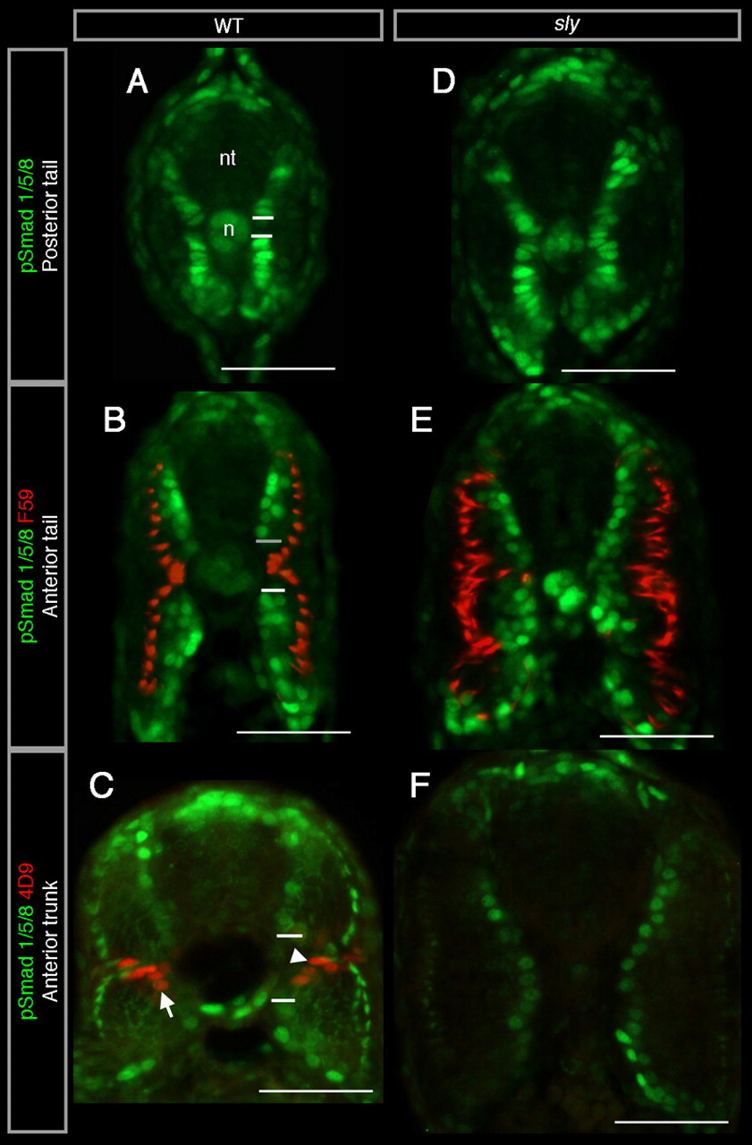
Fig. 4 pSmad expression in MFFs and their precursors is regulated in a similar way to that in pioneers. (A-F) Transverse sections of 26 hpf wild-type (A-C) and sly (D-F) embryos at the level of the posterior tail (A,D), the anterior tail (B,E) and the anterior trunk (C,F). Dorsal is upwards. Nuclear pSmad labelling is in green. Anti-pSmad antibody decorates myofibrils in differentiating slow and fast muscle fibres (C,F). (B,E) F59 (in red) labels migrating slow muscle fibres. (C,F) 4D9 (in red) labels pioneers (arrowhead) and MFFs (arrow). The horizontal white lines delimitate the pSmad-negative central domain. n, notochord; nt, neural tube. Scale bar: 50 μm.