
|
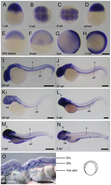
Fig. 5 Spatial and temporal expression of cfl1 during embryogenesis.
(A-N) Representative whole-mount in situ hybridization photographs are shown to reveal the expression patterns of cfl1 at the designated stages from 1-cell to 5 day post-fertilization (dpf) as denoted at the lower left corner of each panel. (O) A representative cryo-section photograph of an embryo at the 70% epiboly stage underwent WISH against cfl1. The cryo-section was taken from the box region as depicted in the embryo carton shown on the right. b, brain; ba, bronchial arches; ll, lateral line system; pd, pronephric duct; pa, pharyngeal arches; EVL, enveloping layer; DEL, deep cell layer. Scale bars: 400 μm for the 4- and 5-dpf embryos, 200 μm for the others and 50 μm for the cryo-section photograph.