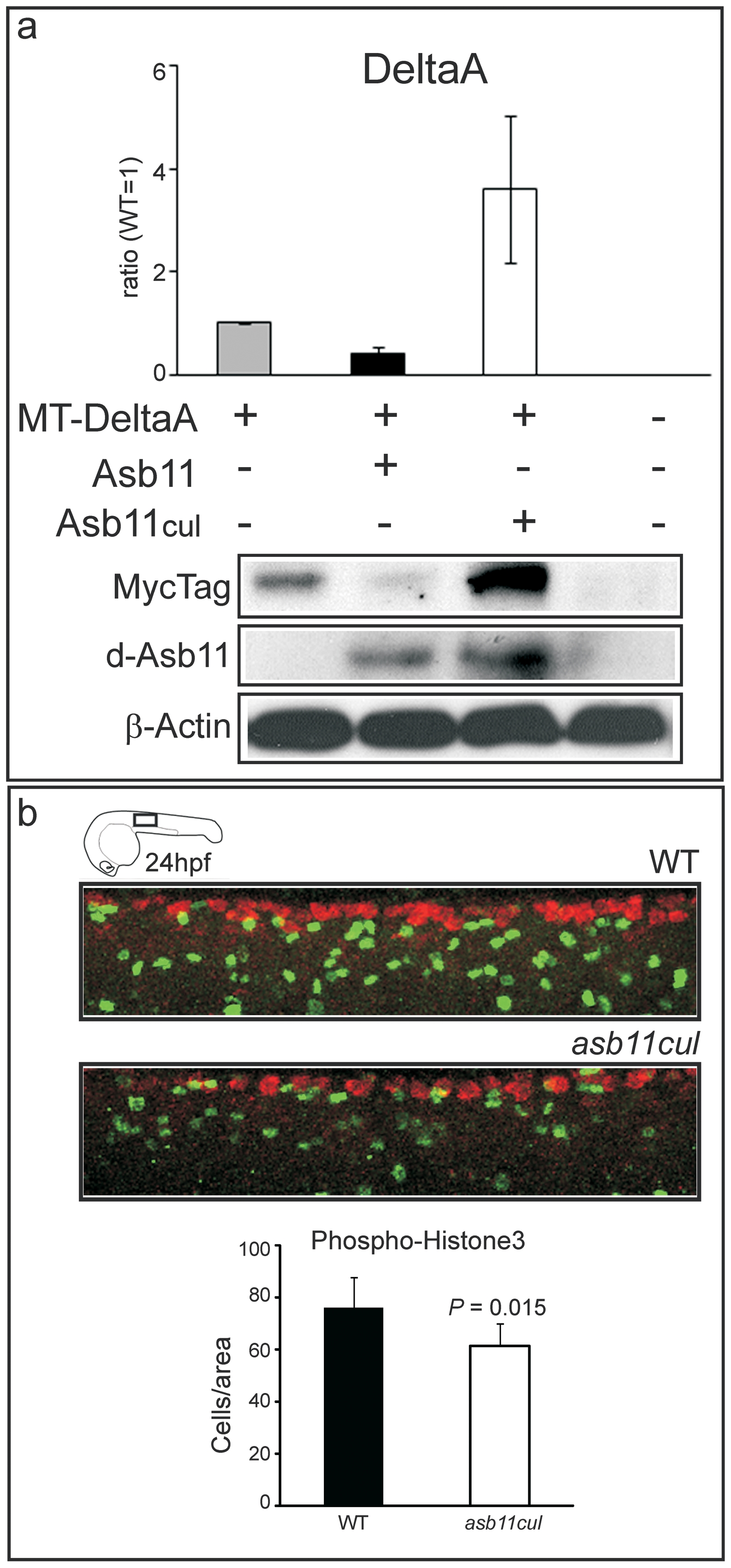
Fig. 6 Cullin box is essential for DeltaA degradation and for maintaining a cell proliferating state in vivo.
(A) Zebrafish embryos were injected with Myc-tagged deltaA (MT-DeltaA) and d-asb11 (Asb11) or asb11cul (Asb11cul) mRNA at one-cell stage. (lower panel) Lysates of 12 hpf embryos were analyzed by immunoblotting for the presence of DeltaA. (higher panel) Graph quantifies 2 individual experiments, each with 30 injected embryos/group. (B), Fluorescent whole-mount antibody labeling of wild type (WT) and asb11cul embryos at 24 hpf for the mitotic marker anti-phosphohistone-3 (PH 3) antibody (green) and the neuronal marker Hu(C). Graph shows the number of positive cells per area (5 somites from beginning of yolk extension) of 5 embryos for each genotype.

