
|
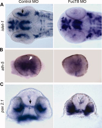
Fig. 4 Patterning defects in the eye and forebrain of 27 hpf FucT8 morphant embryos. A: Ventral view of a FucT8 morphant embryo showing severe loss of islet-1 expressing retinal precursors (arrow). Ninety percent of 20 injected embryos showed this phenotype. B: Whole eyes showing a reduced number of ath-5-positive RGC precursors (arrow) in FucT8 morphant embryos. Similar to eighty-five percent of 40 injected embryos showed this phenotype. C: In situ hybridization using pax 2.1 shows that morphant embryos have a shorter and thicker optic stalk, relative to controls (arrow), and increased pax 2.1 expression in the eye field. Similar to eighty-five percent of 40 injected embryos showed this phenotype.