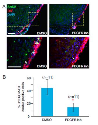
Fig. S6 PDGF signaling is essential for proliferation of epicardial cells during heart regeneration. Treatment of regenerating hearts with DMSO as a control or with PDGFR inhibitor (inh.). The hearts were treated from 2–7 dpa and collected at 7 dpa. (A) Epicardium of zebrafish hearts was labeled with Cell Tracker CMDiI at 2 dpa (BrdU, green; CM-DiI, red). The dashed line marks the approximate position of the amputation plane. BrdU-positive epicardial cells near the wound (marked by arrows) were quantified. (Scale bars = 50 μm.) (B) Statistical analysis of in vivo BrdU incorporation in epicardial cells during heart regeneration. BrdU-positive cells in the boxed area in A were quantified. Eleven hearts (n = 11) were analyzed in each group. Error bars indicate SEM. *P < 0.01.
Image
Figure Caption
Acknowledgments
This image is the copyrighted work of the attributed author or publisher, and
ZFIN has permission only to display this image to its users.
Additional permissions should be obtained from the applicable author or publisher of the image.
Full text @ Proc. Natl. Acad. Sci. USA

