
Image
|
Figure Caption
Fig. S2
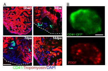
Thrombocytes present at the wound site express pdgfb. (A) Thrombocytes marked by CD41-EGFP fluorescence are localized in the wound area from 3-14 dpa (CD41-GFP, green; DAPI, blue; tropomyosin, red). (Scale bar = 100 μm.) (B) Immunostaining of PDGF-B (red) in a CD41-EGFP-positive thrombocyte. The staining pattern indicated the localization of PDGF-B in α-granules of thrombocytes. (Scale bar = 2.5 μm.)
Figure Data
Acknowledgments
This image is the copyrighted work of the attributed author or publisher, and
ZFIN has permission only to display this image to its users.
Additional permissions should be obtained from the applicable author or publisher of the image.
Full text @ Proc. Natl. Acad. Sci. USA