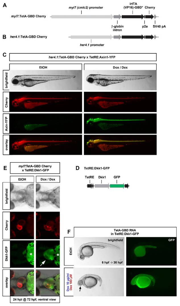
Fig. S3 Conditional, spatially controlled induction using additional TetA-GBD drivers and TetRE responders. (A) Transgenic driver construct for expression of TetA-GBD Cherry specifically in the myocardium. (B) Transgenic driver construct for expression of TetA-GBD Cherry specifically in her4.1 expression domains, primarily in the CNS. (C) her4.1:TetA-GBD Cherry drives efficient induction of Axin1-YFP in embryos doubly transgenic with TetRE:Axin1-YFP. Embryos were treated with EtOH or 25 μg/mL Dox plus 100 μM Dex from 24 h postfertilization (hpf) and photographed at 48 hpf. (D) Transgenic responder line construct for expression of Dkk1-GFP. (E) Heart-specific expression of Dkk1-GFP in myl7:TetA-GBD Cherry; TetRE:Dkk1-GFP double transgenic fish. Images of double transgenic embryos at 72 hpf, treated with EtOH vehicle or 25 μg/mL Dox plus 100 μM Dex from 48 hpf. Note that secreted Dkk1-GFP protein accumulates in the pericardial sac (arrow) in Dox/Dex-treated embryos. *Yolk background fluorescence. n = 20 EtOH, 24 Dox/Dex. (F) Wnt/β-catenin loss-of-function phenotypes as evidenced by posterior truncations and expanded eyes (arrow) in TetRE:Dkk1-GFP embryos injected with 95 pg TetA-GBD RNA and treated with 10 μg/mL Dox plus 100 μM Dex from 4 hpf until 24 hpf. n = 7 EtOH, 8 Dox/Dex.
Image
Figure Caption
Acknowledgments
This image is the copyrighted work of the attributed author or publisher, and
ZFIN has permission only to display this image to its users.
Additional permissions should be obtained from the applicable author or publisher of the image.
Full text @ Proc. Natl. Acad. Sci. USA

