Fig. 5
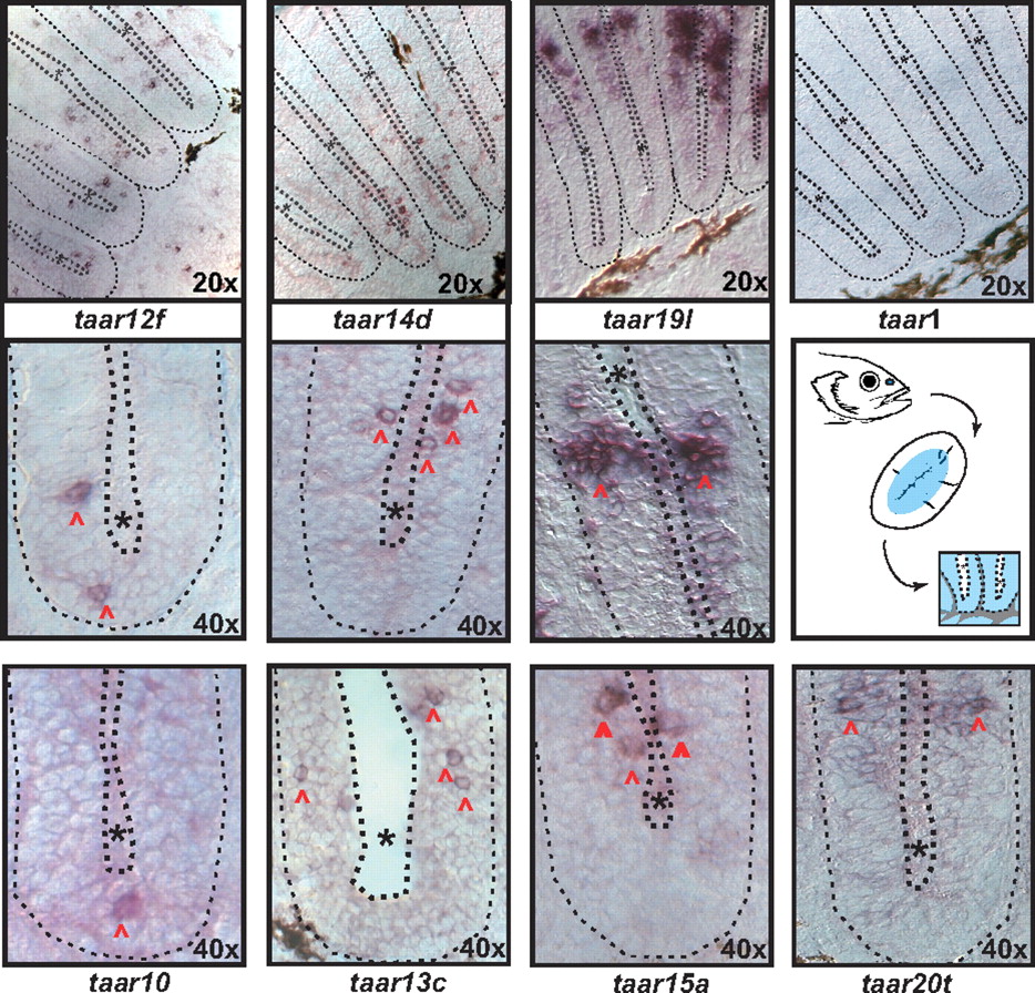
Fig. 5 Expression of taar genes in the zebrafish olfactory epithelium (OE). A schematic representation shows the approximate position of the olfactory epithelium in the zebrafish, the morphology of a horizontal section (lamellae are cut perpendicular to their flat face) and finally an enlargement of 2 lamellae. The central blue-colored area in the lamellae indicates the location of the sensory neuroepithelium (see ref. 20); gray areas and thin dotted line, basal lamina; black dots and asterisk, lumen. In situ hybridization was performed in horizontal sections with antisense RNA probes. The top row depicts the sensory region of several lamellae, whereas the other 2 rows show enlargements of 1 lamella, corresponding approximately to one-half of the schematical representation (Center Right). Red arrowheads point to labeled neurons, other symbols as above. Taar genes 10, 12f, 13c, 14d, and 15a are expressed in sparse cells, whereas taar 19l and 20t label a somewhat larger subset of cells within the sensory surface, probably because of cross-hybridization in the large and closely related subfamilies taar 19 and taar 20.

