
|
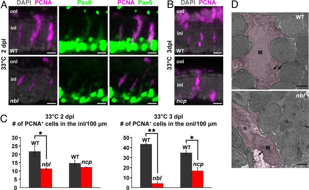
Fig. 4
Retinal regeneration defects of nbl and ncp. (A) Neurogenic clusters at 2 dpl in the inner nuclear layer (inl) immunolabeled with anti-PCNA (magenta) and weakly labeled with anti-Pax6 (green) in WT and nbl. Note that Pax6 is also expressed at high levels in amacrine cells at the inner boundary of the inl. (B) PNCA+ photoreceptor progenitors at 3 dpl in the outer nuclear layer (onl) of WT and ncp. (C) Number of PNCA+ cells in the inl or onl per 100 μm of linear length retina at 2 or 3 dpl, respectively. Error bars represent SEM for 3 individuals. *, P < 0.05; **, P < 0.0001. (D) Transmission electron micrographs of injury-activated M�ller glia in WT and nbl. See text for description of temperature shift paradigm. M�ller glia (M) are shown by the magenta wash. Mitochondria (arrows) in M�ller glia of WT appear normal after 8 h at 33�C, whereas in nbl mutants, M�ller glia contain swollen mitochondria. (Scale bars: 10 μm in A and B; 100 μm in D.)