
|
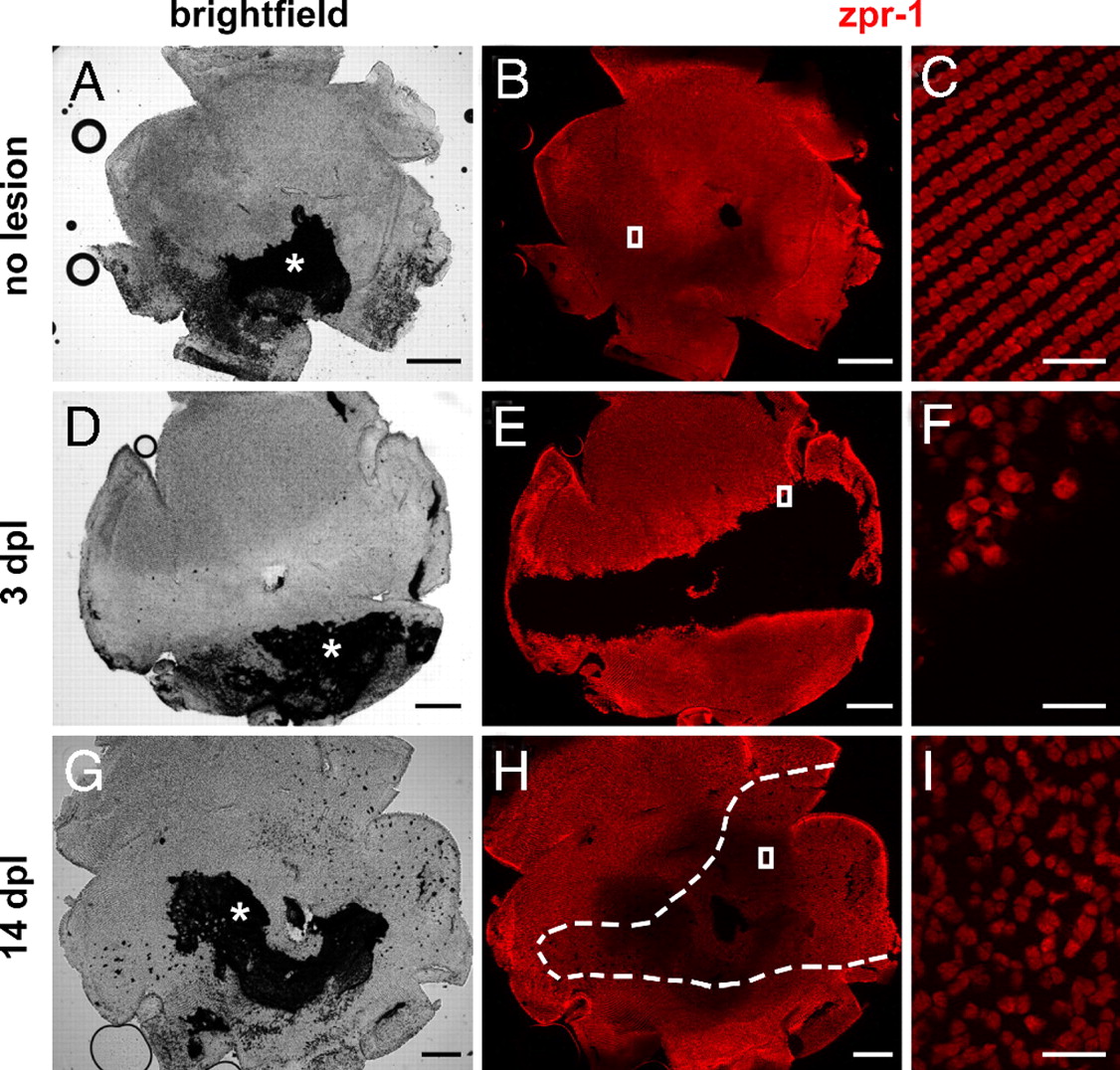
Fig. 1
Cone photoreceptor regeneration in adult zebrafish. Flat-mounted zebrafish retinas are immunolabeled with cone-specific zpr-1 (red). Retinas are oriented dorsal up, ventral down, nasal left, and temporal right. (A, B) Intact retina. Asterisk, attached retinal pigment epithelium. (D, E) At 3 days after exposure to intense light, cones are missing in a horizontal band across the retina. (G, H) By 14 days, cones have regenerated within the lesioned region (dashed lines). (C, F, and I) Magnified images of the boxes in B, E, and H, respectively. (Scale bars: 300 μm in A, B, D, E, G, and H; 20 μm in C, F, and I.)