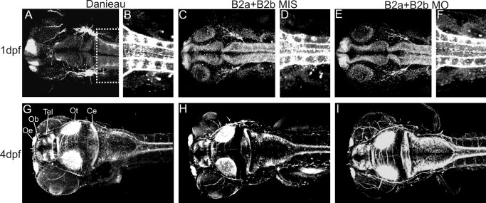
Fig. 7 Whole-mount staining with anti-acetylated α-tubulin antibody to view axon projections in 1 days postfertilization (dpf) and 4 dpf zebrafish. A-F: Confocal scans of 1dpf zebrafish embryos. A: A dorsal view of the head of a Danieau-treated embryo. B: A ventral view of the spinal axons (area boxed in A). C-F: B2aMIS+B2bMIS-treated (5 ng+5 ng, C,D) and B2aMO+B2BMO-treated (5 ng+5 ng, E,F) embryos. G?I: Ventral stacks of 4 dpf zebrafish embryo head after Danieau-treatment (G), B2a+B2bMIS treatment (H), and B2aMO+B2bMO treatment (I). Ce, cerebellum; Ob, olfactory bulb; Oe, olfactory epithelium; Ot, optic tectum; Tel, telencephalon.
Image
Figure Caption
Acknowledgments
This image is the copyrighted work of the attributed author or publisher, and
ZFIN has permission only to display this image to its users.
Additional permissions should be obtained from the applicable author or publisher of the image.
Full text @ Dev. Dyn.

