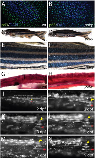
Fig. 7 poky mutant adults have normal skin and wild type EVL cells persist into larval stages. (A,B) Optical section through wild type (A) and poky mutant (B) epidermis expressing nuclear p63 protein (green) at 24 hpf. (C,D) 2 year-old wild type and poky mutants have no obvious skin lesions. (E,F) Pigment, scales and fins are normal in wild type (E) and poky mutants (F). Histology of adult skin reveals the typical bilayered epidermis (bracket) in wild type (G) and poky mutants (H). Lineage tracing of embryonic and larval EVL (I?N). EVL from embryos scatter labeled with fluorescent dextran was observed up to 9 dpf. (I) 2 dpf low magnification image of the tail shows multiple cells labeled; location at higher magnification in J. (J) A clone of EVL cells is easily observable at 2 dpf (yellow arrowhead marks a single EVL cell). The location of these cells was traced relative to underlying muscle cells (red asterisk) at 2 dpf (J), 3 dpf (K), 5 dpf (L), 7 dpf (M) and 9 dpf (N).
Reprinted from Developmental Biology, 346(2), Fukazawa, C., Santiago, C., Park, K.M., Deery, W.J., Canny, S.G., Holterhoff, C.K., and Wagner, D.S., poky/chuk/ikk1 is required for differentiation of the zebrafish embryonic epidermis, 272-283, Copyright (2010) with permission from Elsevier. Full text @ Dev. Biol.

