
|
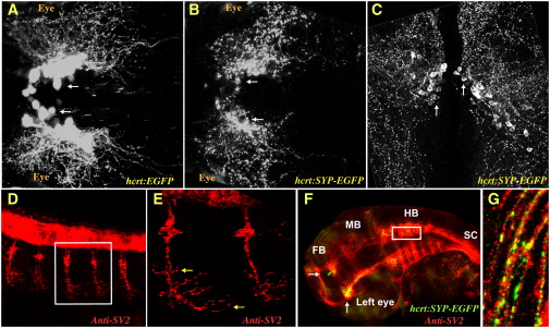
Fig. 1 The Presynaptic SYP-EGFP Fusion Protein Is a Marker of Synapses in HCRT Axons
(A and B) Two-photon microscope imaging of transgenic larvae (dorsal views of the midbrain, anterior to the left) expressing EGFP (hcrt:EGFP, A) in all HCRT neurons or the presynaptic marker synaptophysin fused to EGFP (hcrt:SYP-EGFP, B) to specifically target synapses in the axons of HCRT neurons.
(C) Confocal imaging of 100 μm transversal brain sections from a stable hcrt:SYP-EGFP transgenic adult fish. HCRT cell bodies are localized around the ventricle in the hypothalamus and SYP-EGFP puncta are distributed throughout HCRT axons.
(D?G) Confocal imaging (lateral views with anterior to the left) of immunohistochemistry (with SV2 antibody, D and E) and double-immunohistochemistry (with SV2 and EGFP antibodies, F and G) in a 32 hr postfertilization (hpf) embryo. (E) and (G) are close-ups of the white frames of (D) and (F), respectively. Of note, red (SV2) and green (SYP-EGFP) colocalization is marked with yellow. The following abbreviations are used: FB, forebrain; MB, midbrain; HB, hindbrain; and SC, spinal cord. White and yellow arrows indicate HCRT cell bodies and SV2 presynaptic clusters in motor neurons, respectively.