
|
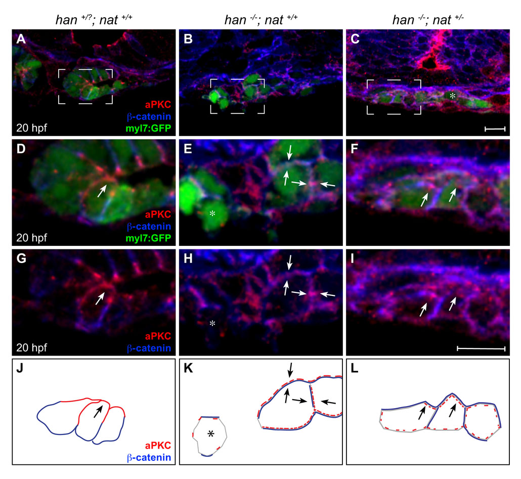
Fig. S4 Poor rescue of myocardial apicobasal polarity in han-/-;nat+/- embryos. (A-I) Transverse confocal sections of the right lateral mesoderm in embryos expressing Tg(myl7:egfp) (green). Dorsal is up. Immunofluorescence detects aPKC (red) and β-catenin (blue). Boxed regions in A-C are shown at higher magnification in D-I. At this stage, the wild-type myocardium has begun the asymmetric involution that displaces a medial portion of the myocardial epithelium dorsally (Rohr et al., 2008). Scale bars: 10 μm. (J-L) Cartoons schematize cardiomyocyte-associated localization of aPKC (red) and β-catenin (blue) shown in D-I. Wild-type cardiomyocytes are organized in a cohesive layer (A) and exhibit apical enrichment of aPKC (D,G,J, arrow) and basolateral enrichment of β-catenin (D,G,J). In han embryos, the small clusters of cardiomyocytes (B) exhibit irregular localization of aPKC and β-catenin, either diffusely around the cell (E,H,K, arrows) or nearly absent (E,H,K, asterisk). Only slight improvements are seen in han-/-;nat+/- embryos: aPKC is either distributed diffusely (C, asterisk) or partially enriched apically (F,I,L, arrows), and β-catenin is either distributed diffusely or partially enriched laterally (F,I,L). We note that previous work indicated an absence of aPKC localization in han mutant cardiomyocytes (Trinh et al., 2005). We suspect that the diffuse and irregular aPKC localization observed here (E,H,K) reflects increased sensitivity of our current protocol, which uses a different fixation method and thinner sections.