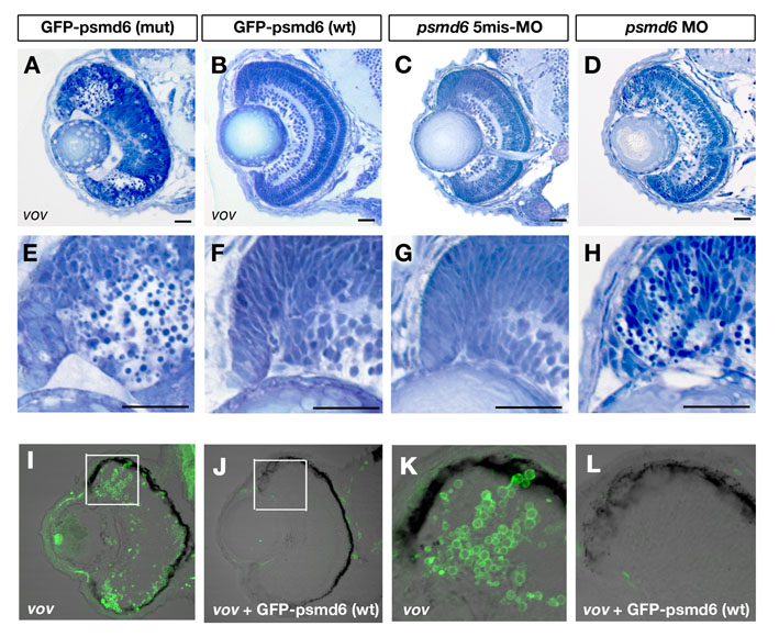
Fig. S8 Retinal phenotypes of vov mutants injected with GFP-tagged wild-type and vov mutant form psmd6 RNA and of psmd6 morphant. (A,B) Plastic sections of vov mutants injected with GFP-tagged wild-type (A) and vov mutant form psmd6 RNA (B) at 72 hpf. (E,F) Higher magnification of retinal CMZ of A,B. Pyknotic nuclei are observed in the neurogenic region of retinal CMZ of the vov mutant injected with GFP-tagged psmd6 mutant form RNA. By contrast, apoptosis in retinal CMZ is suppressed and retinal lamination is normal in the vov mutant injected with GFP-tagged wild-type psmd6 RNA. (C,D) Plastic sections of retinas of psmd6 five-mismatch morphant (C) and psmd6 morphant (D) at 72 hpf. (G,H) Higher magnification of retinal CMZ of C,D. Apoptosis is observed in retinal CMZ of psmd6 morphant (D), but not in psmd6 five-mismatch morphant (C). (I-L) TUNEL of vov mutant (I,K) and vov mutant injected with wild-type psmd6 RNA (J,L). (K,L) High-magnification images of boxed regions in I,J. The number of TUNEL-positive cells in the retinal CMZ is significantly reduced in the vov mutant injected with psmd6 RNA as compared with the vov mutant. Scale bars: 50 μm.
Image
Figure Caption
Acknowledgments
This image is the copyrighted work of the attributed author or publisher, and
ZFIN has permission only to display this image to its users.
Additional permissions should be obtained from the applicable author or publisher of the image.
Full text @ Development

