Image
Figure Caption
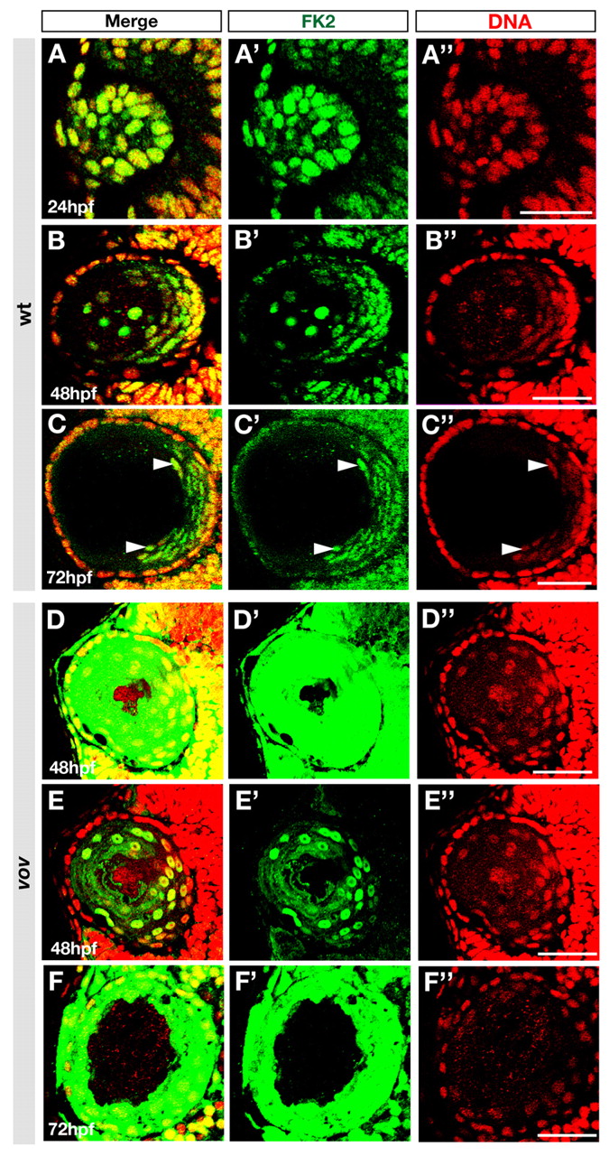
Fig. 5 Polyubiquitylated proteins are localized in lens fiber cell nuclei. (A-F) Labeling of wild-type (A-C) and vov mutant (D-F) zebrafish lens of the indicated stages with anti-polyubiquitylated protein antibody (FK2, green) and TOPRO3 (red). Arrowheads indicate signals in nuclei of differentiating lens fiber cells at 72 hpf (C). Scanning conditions of images in D,F were the same as those for B,C. (E) The same image as D but with reduced sensitivity of fluorescence detection. (A′-F′,A"-F") The same images as A-F but shown through a green (FK2) or red (TOPRO3) channel. Scale bars: 50 Ám.
Figure Data
Acknowledgments
This image is the copyrighted work of the attributed author or publisher, and
ZFIN has permission only to display this image to its users.
Additional permissions should be obtained from the applicable author or publisher of the image.
Full text @ Development

