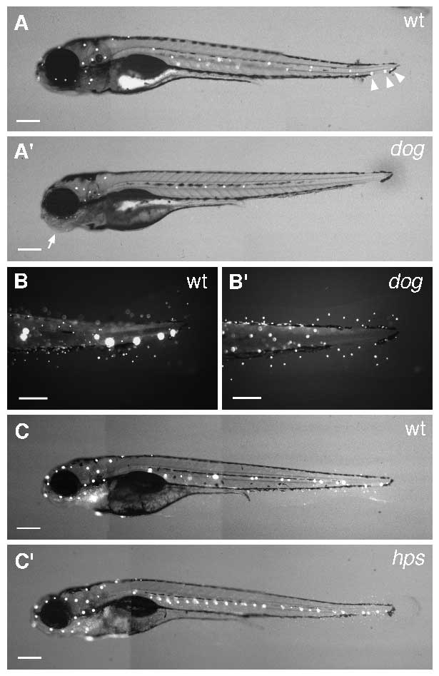
Fig. 6 DASPEI stain of lateral line neuromasts at 120 hours. (A,A′) Wild-type (wt) and dog embryo. Neuromasts appear as bright dots; their distribution is highly consistent between wild-type individuals (compare A and C). dog lacks the set of neuromasts at the tail tip (arrowheads in wild type). Note the abnormal ear and jaw morphology (arrow) of the dog embryo. Scale bar, 200 μm. (B,B′) Higher magnification of the tail of wt and dog embryos. Individual cells on the skin (identity unknown) that also stain with DASPEI are evident in dog, but neuromasts (larger dots) are absent. Scale bar, 100 μm. (C,C′) Comparison of neuromast pattern in wt and hps embryos. Note the extra neuromasts in the trunk of the mutant. Scale bar, 200 μm.
Image
Figure Caption
Figure Data
Acknowledgments
This image is the copyrighted work of the attributed author or publisher, and
ZFIN has permission only to display this image to its users.
Additional permissions should be obtained from the applicable author or publisher of the image.
Full text @ Development

