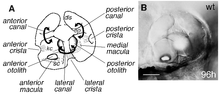
Fig. 1 Structural features of the wild-type zebrafish ear. Anterior is to the left in all Figs. (A) Cut-away lateral view of a wild-type zebrafish ear at 96 hours, to give a three-dimensional impression of the structures within the vesicle. (B) Photograph of an ear at the same age, taken with differential interference contrast (DIC) optics, and focussed at the level of the anterior otolith. Note the overall shape of the organ. Epithelial projections (ep) within the ear form hubs of the developing semicircular canals (curved arrows). Each canal (anterior, lateral and posterior) is associated with a small sensory patch or crista, while each otolith overlies a larger sensory patch or macula. Kinocilia of the crista hair cells (kc) are long, and project into the canal lumens. Kinocilia of the maculae are shorter and the otoliths appear to sit directly on the stereociliary bundles (sc) of the macular hair cells. The smaller (anterior) otolith lies in a lateral position; the larger (posterior) otolith lies medially. dls, dorsolateral septum. Scale bar, 50 μm.
Image
Figure Caption
Acknowledgments
This image is the copyrighted work of the attributed author or publisher, and
ZFIN has permission only to display this image to its users.
Additional permissions should be obtained from the applicable author or publisher of the image.
Full text @ Development

