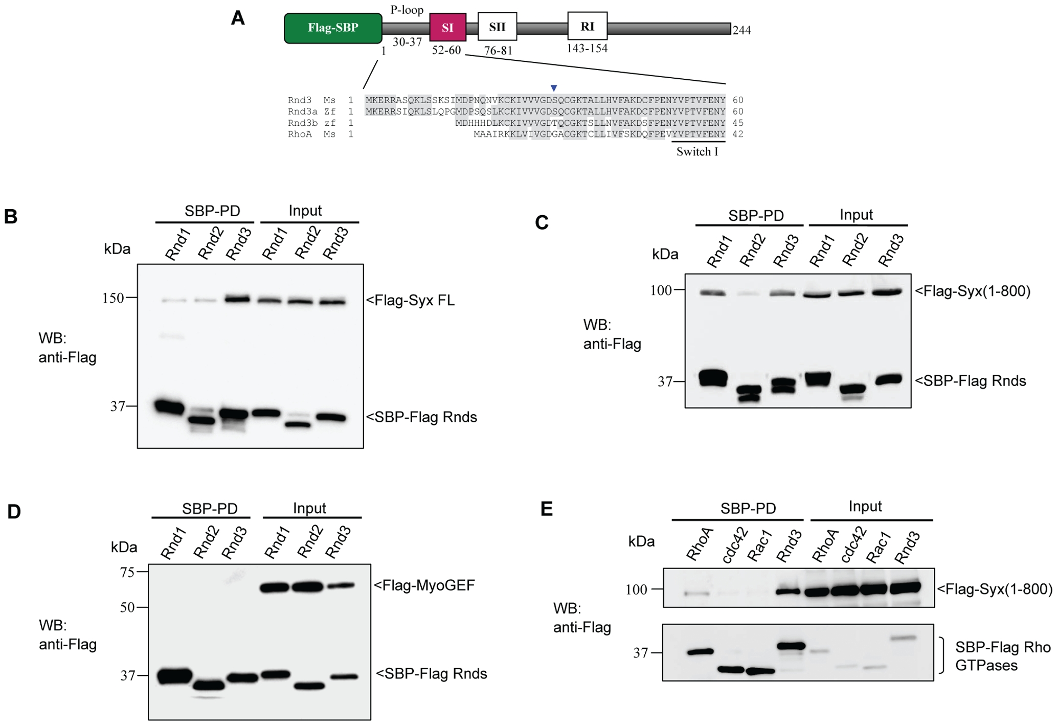
Fig. 2 Analysis of the Rnd3-Syx interaction.
(A) Schematic representation of Flag-SBP tagged Rnd3. The motifs of Rho family proteins are indicated: P-loop, the switch I and II (SI and SII), and Rho insert (RI) region. Flag (FL) and streptavidin binding peptide (SBP) tags are indicated. Sequence alignment of Rnd3 proteins showed that the N-terminal of zebrafish Rnd3a resembles more closely to mouse Rnd3. Identical residues are shaded in grey. Blue arrowhead denotes G14 in RhoA. Residue effector region is underlined. The accession numbers of mouse Rnd3 (NP083086), zebrafish Rnd3a (BC059452), Rnd3b (BC076009) and mouse RhoA (NP058082) are in brackets. (B and C) Characterization of Syx binding to Rnd isoforms. 293T cells were co-transfected with expression vectors encoding SBP-Flag Rnd1, Rnd2 or Rnd3 and full-length Syx or Syx(1?800) as indicated. Lysates were subjected to SBP pulldown and immunoblotting. (D) Rnd3 does not interact with MyoGEF. 293T cells were co-transfected as for (B) with Flag-MyoGEF and assessed for interaction. (E) Selective association of Syx with Rnd3. Co-expression of SBP-Flag RhoA(GV14), Cdc42(G12V), Rac1(G12V) or Rnd3 with Syx(1?800) and immunoblotted for Flag epitope.

