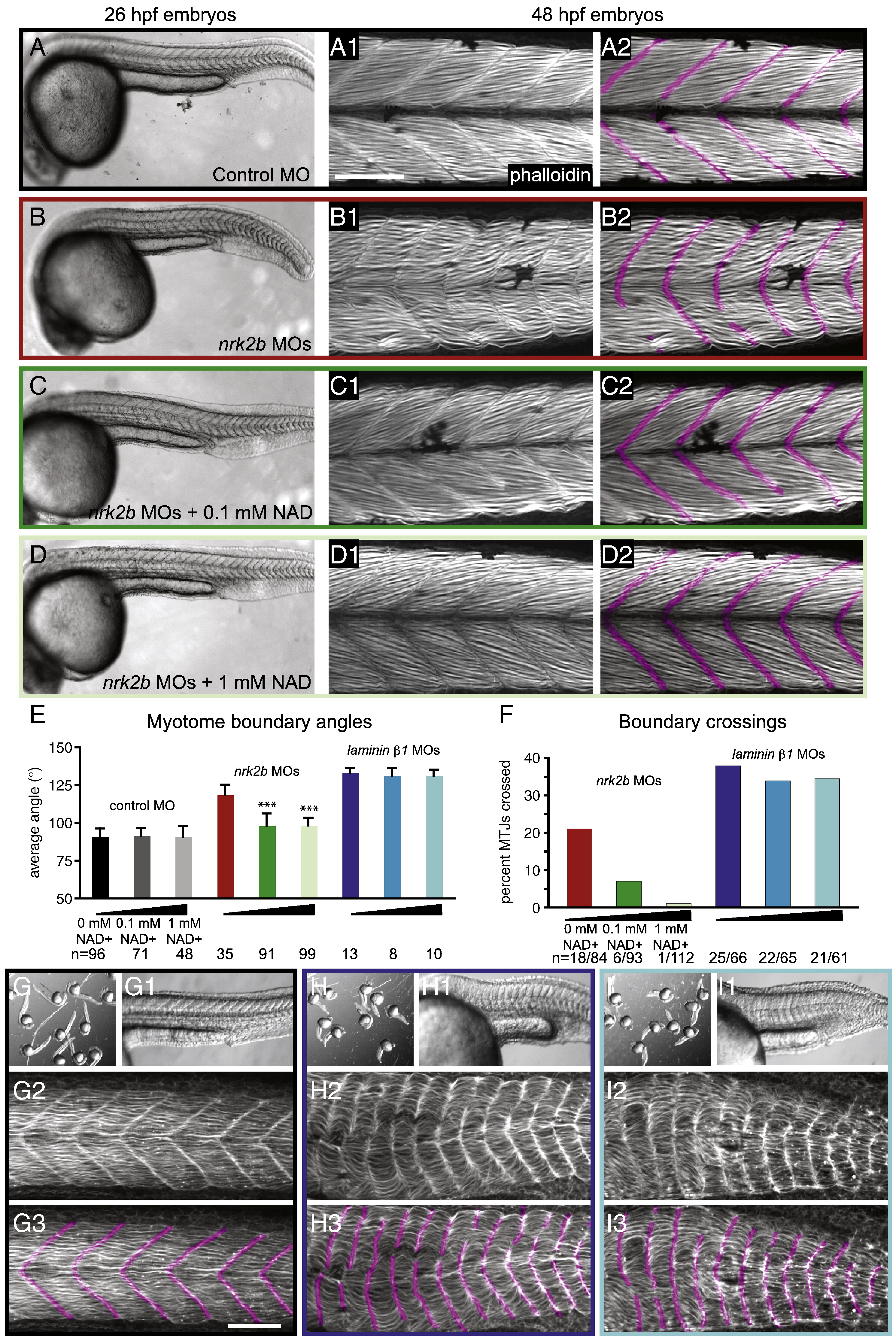
Fig. 3 Exogenous NAD+ treatment rescues the nrk2b morphant muscle phenotype. (A–D) Brightfield images, side mount, anterior left, dorsal top, 26 hpf embryos. (Numbered panels A–D) Side mount, anterior left, dorsal top, 48 hpf embryos, phalloidin stained to visualize actin. In panels numbered 2, MTJs are pseudocolored fuchsia. (Panel A) Control MO-injected. (Panel B) nrk2b MO-injected. (Panel C) nrk2b MO-injected embryos incubated in 0.1 mM NAD+. (Panel D) nrk2b MO-injected embryos incubated in 1 mM NAD+. (E) Graph of average myotome boundary angles in controls, NAD+-treated controls, nrk2b morphants, NAD+-treated nrk2b morphants, laminin morphants, and NAD+-treated laminin morphants. The average myotome boundary angle of nrk2b morphants is significantly different than controls (Student t-test, p < 0.001) and the average myotome boundary angle of NAD+-treated nrk2b morphants is significantly different than untreated nrk2b morphants and controls (Student t-test, p < 0.001, n of myotome boundaries measured on x-axis). (F) Graph of the percent of MTJs crossed by muscle fibers in nrk2b morphants, NAD+-treated nrk2b morphants, laminin morphants, and NAD+-treated laminin morphants. NAD+ treatment rescues boundary crossings in a dose dependent manner in nrk2b morphants, but not laminin morphants (n of MTJs crossed over total MTJs analyzed is listed on the x-axis). (Panel G) Controls. (Panel H) laminin morphants. (Panel I) 1 mM NAD+-treated laminin morphants. (Lettered panels) Brightfield images of a dish of embryos. (Panels numbered 1) Side mount, brightfield images, 26 hpf embryos, anterior left, dorsal top. (Panels numbered 2) Side mount, 48 hpf embryos, anterior left, dorsal top, phalloidin stained to visualize actin. (Panels numbered 3) MTJs are pseudocolored fuchsia. Scale bars are 50 μm, ***p < 0.001.
Reprinted from Developmental Biology, 344(2), Goody, M.F., Kelly, M.W., Lessard, K.N., Khalil, A., and Henry, C.A., Nrk2b-mediated NAD+ production regulates cell adhesion and is required for muscle morphogenesis in vivo: Nrk2b and NAD+ in muscle morphogenesis, 809-826, Copyright (2010) with permission from Elsevier. Full text @ Dev. Biol.

