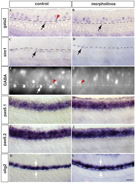
Fig. S1 Lateral floor plate identity depends on nkx2.2a, nkx2.2b and nkx2.9. (A,B) Control (A) and Mo-nkx2.2a/Mo-nkx2.2b/Mo-nkx2.9-injected embryo (B) stained with gata2 antisense probe. Triple knockdown abolished gata2-positive cells in the lateral floor plate (arrow) completely. gata2-expressing cells (arrowhead) located dorsally in the spinal cord were unaffected. (C,D) Control (C) and nkx2 triple-knockdown (D) embryos hybridized to sim1 antisense probe. Knockdown of all three Nkx2 genes deleted sim1 expression in the lateral floor plate (arrow). (E,F) Control (E) and triple-knockdown (F) embryos labeled with anti-GABA antibody. Anti-GABA staining was completely lost in the lateral floor plate (arrow) in the triple-knockdown embryo. GABAergic neurons (arrowhead), which are located more dorsally, are not affected by knockdown of the three Nkx2 genes. (G,H) Control (G) and triple-knockdown (H) embryos stained with pax6.1 antisense probe. The expression of pax6.1 is not affected by the triple knockdown. (I,J) Control (I) and triple-knockdown (J) embryos stained with pax6.2 probe. As in the case of pax6.1, pax6.2 is not expanded into the lateral floor plate in Nkx2 triple-knockdown embryos. (K,L) Control (K) and triple-knockdown (L) embryo hybridized to olig2 probe. olig2-positive cells are found ectopically in the lateral floor plate in nkx2 triple-knockdown embryos. The dashed line indicates the border between the lateral floor plate and more dorsally located cells. Arrowheads outline the increased width of the olig2 expression domain in the morphants. pax6.1 and pax6.2 expression does not expand into the lateral floor plate in triple-knockdown embryos, whereas olig2-expressing cells were aberrantly found in the lateral floor plate in these knockdown embryos. Representative lateral views of the spinal cord over the hindgut extension are shown. Embryos are 48 hpf (C,D) and 24 hpf (A,B,E-L) and are oriented with anterior left and dorsal up. Scale bar: 25 μm.
Image
Figure Caption
Acknowledgments
This image is the copyrighted work of the attributed author or publisher, and
ZFIN has permission only to display this image to its users.
Additional permissions should be obtained from the applicable author or publisher of the image.
Full text @ Development

