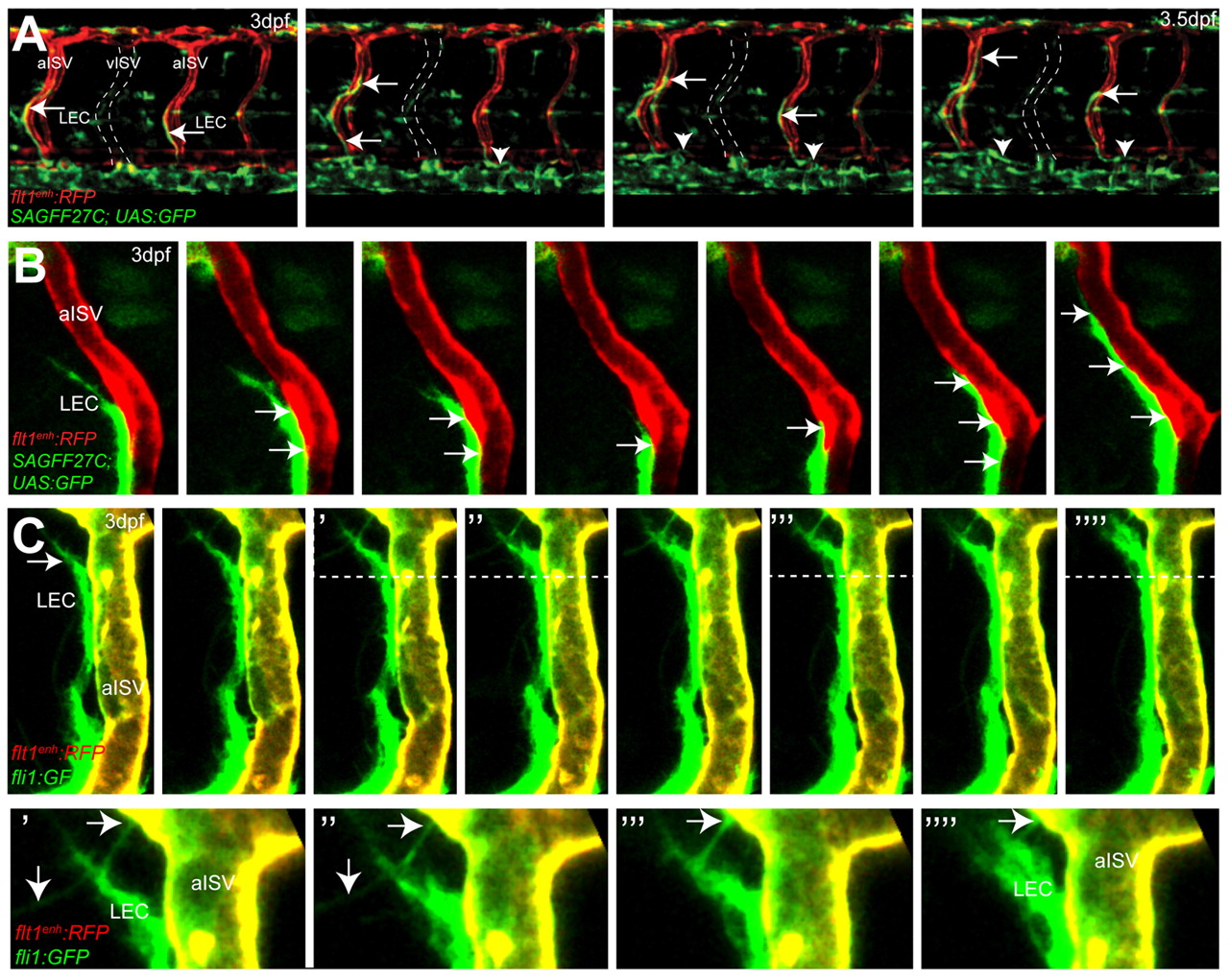
Fig. 3 Migration of lymphatic precursors along intersegmental arteries. (A) Still images of flt1enh:RFP;SAGFF27C;UAS:GFP triple transgenic embryos reveal migration of SAGFF27C;UAS:GFP+ LECs (arrows) along flt1enh:RFP+ arteries and establishing the TD (arrowheads) in the trunk. The position of a vISV is indicated by broken lines. (B) Still images of migrating SAGFF27C;UAS:GFP+ LECs suggest direct contact (arrows) between the aISV and the LEC. (C) Still images of migrating fli1a:GFP+ LECs and filopodia formation (indicated by arrows) at the leading edge along the surface of a flt1enh:RFP+; fli1a:GFP+ artery at 3 dpf. Areas above the broken lines are shown at higher magnification in the bottom row.
Image
Figure Caption
Figure Data
Acknowledgments
This image is the copyrighted work of the attributed author or publisher, and
ZFIN has permission only to display this image to its users.
Additional permissions should be obtained from the applicable author or publisher of the image.
Full text @ Development

