
|
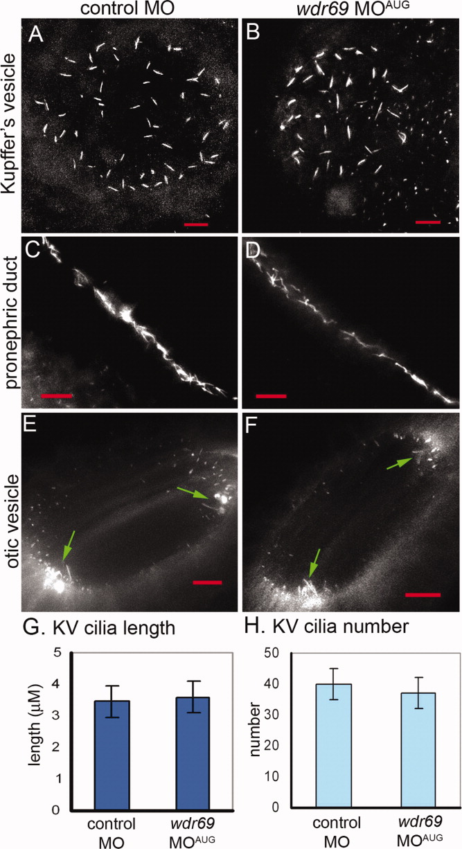
Fig. 4 Cilia length and number is not changed in wdr69 morphants. A-F: Cilia were visualized by immunofluorescent staining with acetylated tubulin antibodies. Cilia in embryos injected with wdr69 MOAUG appeared similar to cilia injected with control MO in Kupffer′s vesicle (KV; A,B), pronephric ducts (C,D) and otic vesicles (arrows in E,F point to motile tether cilia). All red scale bars represent 10 μM. G,H: Measurements of KV cilia at six to eight somite stage (SS) showed no significant differences in the average length (G) or number (H) between wdr69 MOAUG (n = 17) and control (n = 21) morphants. Error bars = one standard deviation. MO, morpholino oligonucleotides.