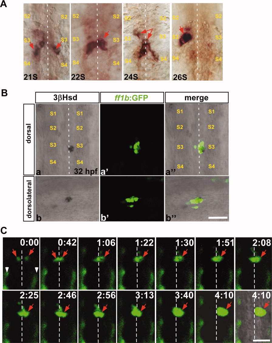
Fig. 1 The ff1b-expressing interrenal primordia during midline fusion and lateral repositioning. A: Ventral flat mount views of 21-somite (21S), 22-somite (22S), 24-somite (24S), and 26-somite (26S) stage embryos, which were subject to ISH for detecting ff1b mRNA, with anterior oriented to the top. B: The colocalization of steroidogenic activity and ff1b:GFP transgene expression at the interrenal tissue, in the Tg(ff1bEx2:GFP) embryo. Confocal images display the steroidogenic cells as detected by 3&geta;Hsd activity staining (a,b), and the green fluorescence driven by ff1b promoter (a′,b′), in a Tg(ff1bEx2:GFP) embryo at 32 hpf. The merged images of 3 μ-Hsd activity staining and GFP are shown in a″,b′. a-a″: Dorsal views with anterior to the top; (b-b″) dorsolateral views with anterior to the right. C: Confocal time-lapse imaging of the interrenal tissue in a live Tg(ff1bEx2:GFP) embryo. A dechorionated embryo at around 21-somite stages was mounted with the dorsal side up in 3% methyl cellulose. The fluorescent images were collected at 1-min intervals, and representative frames are shown. The last frame of the time series is a merge of fluorescent and bright-field images. Time is indicated by hours:minutes. Since the sample was kept at 23°C during observation, developmental stages cannot be accurately addressed. Each fluorescent image in B and C represents a projection of a consecutive z-stack encompassing the depth of the interrenal tissue. Both ISH and time-lapse ff1b:GFP analyses show that the interrenal primordia fuse at the midline prior to the lateral relocalization. Red arrows indicate ff1b-expressing interrenal primordia in A, and the ff1b promoter-driven interrenal-specific fluorescence in C. S2, S3, and S4, the second, third, and fourth somite, respectively. White arrowheads in C indicate ectopic GFP expression in muscle pioneer cells. White dotted lines indicate the position of the midline. Scale bar = 50 μM.
Image
Figure Caption
Figure Data
Acknowledgments
This image is the copyrighted work of the attributed author or publisher, and
ZFIN has permission only to display this image to its users.
Additional permissions should be obtained from the applicable author or publisher of the image.
Full text @ Dev. Dyn.

