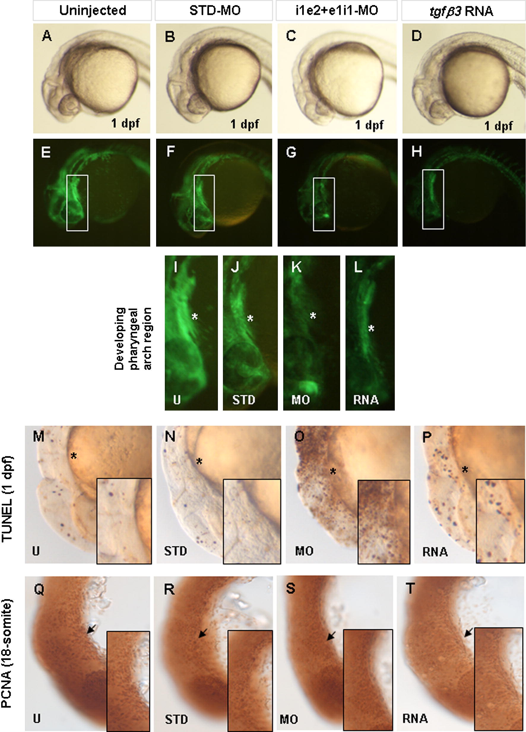
Fig. 7 Effect of loss and over-expression of tgfβ3 on apoptosis and cellular proliferation. Whole-mount lateral (A?P) and flat-mount lateral (Q?T) views are shown in brightfield (A?D, M?T) and darkfield (E?L). The fli1a:gfp transgene is highly expressed in CNC-derived cells of the developing pharyngeal arch of uninjected (A, E, I) and STD-MO injected (B, F, J) transgenic 1 dpf embryos (asterisk). Injection of i1e2 and e1i1-MOs (C, G, K) or tgfβ3 RNA (D, H, L) leads to a visible reduction in transgene expressing cells. TUNEL analysis reveals a marked increase in apoptotic cells in double knockdown embryos (O) compared with uninjected (M) or STD-MO injected (N) embryos, while over-expressing embryos (P) showed a slight increase in apoptotic cells. PCNA immunohistochemistry of 18-somite stage embryos shows similar numbers of PCNA positive cells (arrow) in uninjected (Q), STD-MO injected (R), double knockdown morphant (S), and over-expressing (T) embryos. Insets in M?T show the zoom-in view of the developing pharyngeal arch region.
Reprinted from Mechanisms of Development, 128(7-8), Cheah, F.S., Winkler, C., Jabs, E.W., and Chong, S.S., tgfbeta3 Regulation of Chondrogenesis and Osteogenesis in Zebrafish is Mediated Through Formation and Survival of a Subpopulation of the Cranial Neural Crest, 329-344, Copyright (2010) with permission from Elsevier. Full text @ Mech. Dev.

