
Image
|
Figure Caption
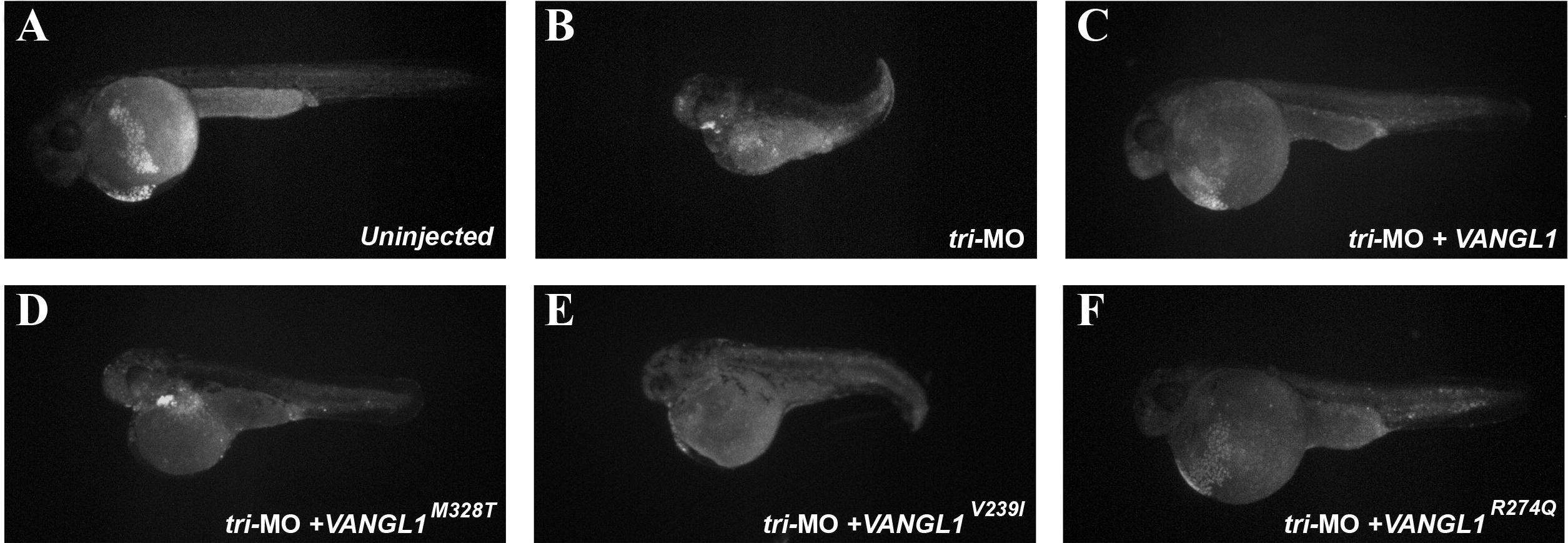
Fig. S2 Little apoptosis in zebrafish lacking tri. Apoptosis was assessed by visualizing acridine orange labeled cells. In wild-type larvae, the majority of dying cells were observed in the yolk (Panel A). In tri-MO injected embryos, there were slightly more labeled cells in the body and the yolk of the larvae (Panels B?F).
Acknowledgments
This image is the copyrighted work of the attributed author or publisher, and
ZFIN has permission only to display this image to its users.
Additional permissions should be obtained from the applicable author or publisher of the image.
Reprinted from Mechanisms of Development, 127(7-8), Reynolds, A., McDearmid, J.R., Lachance, S., Marco, P.D., Merello, E., Capra, V., Gros, P., Drapeau, P., and Kibar, Z., VANGL1 rare variants associated with neural tube defects affect convergent extension in zebrafish, 385-392, Copyright (2010) with permission from Elsevier. Full text @ Mech. Dev.