
|
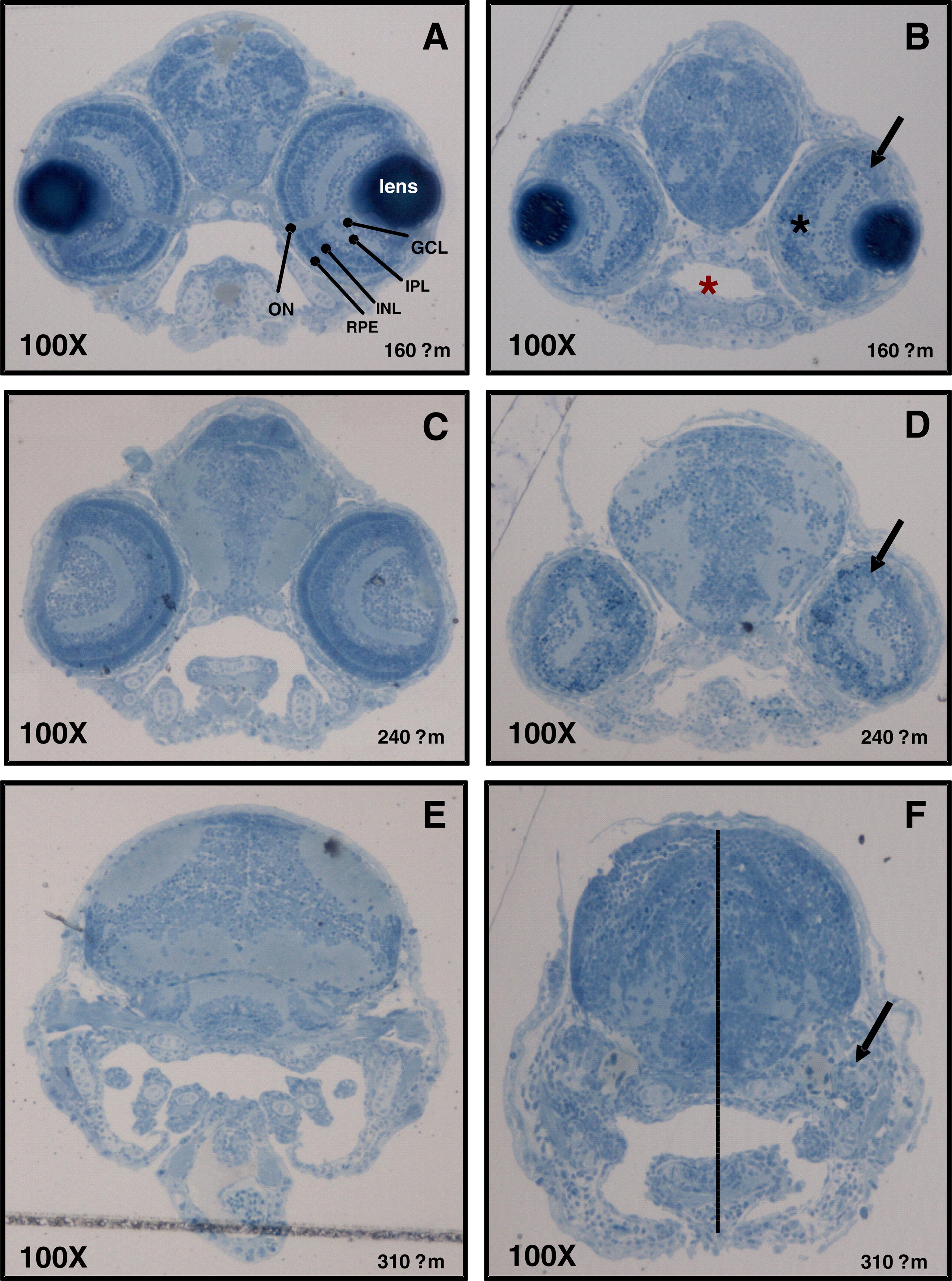
Fig. 5 Characterisation of developmental defects associated with zCactin knockdown. The control morpholino (A, C and E) and Cactin morpholino embryos (B, D and F) at 72 hpf were processed for light microscopy sectioning. Sections at depths of 160, 240, 310 μm into the anterior embryo were obtained using a diamond knife mounted on a Leica EMUC6 ultramicrotome. For histological examination the sections were stained lightly with 1% toludine blue. Distinct layers stratify ganglion, amacrine, bipolar, horizontal and Muller cells as well as rod and cone photoreceptors cells (A). ON, optic nerve; GCL, ganglion cell layer; IPL, inner plexiform layer; INL, inner nuclear layer; RPE, retinal pigmented epithelium. Black arrows indicate the dysmorphic layer of the eye (panel C, F and I). Red asterisk shows the smaller oral cavity in the morphant section (panel C), while black asterisk indicates the more spherical shape of the eye (panel C). Dotted line shows the reduced distance between the crown and the mandibular arch (panel F). Microscopical images were captured using a Leica DM LB microscope equipped with a Leica DFC 480 digital camera.
Reprinted from Gene expression patterns : GEP, 10(4-5), Atzei, P., Yang, F., Collery, R., Kennedy, B.N., and Moynagh, P.N., Characterisation of expression patterns and functional role of Cactin in early zebrafish development, 199-206, Copyright (2010) with permission from Elsevier. Full text @ Gene Expr. Patterns