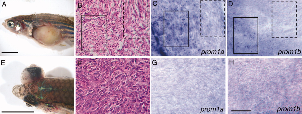
Fig. 5 Increased expression of prom1a and prom1b in epitheloid-like cells in tp53M214K/tp53M214K induced neoplasm. A, B, E, F: Gross morphology and histopathology of abdominal (A, B) and ocular (E, F) neoplasms in 12-month-old adult homozygous tp53M214K/tp53M214K zebrafish. B: Histopathological features of the abdominal neoplasm show regions with predominantly spindle cells (solid line box) or predominantly epitheloid cells (dashed line box), consistent with a diagnosis of zMPNST (Berghmans et al., [2005]). F: Histopathological features of ocular tumor show predominantly spindle cells, consistent with a diagnosis of zMPNST (Berghmans, et al., 2005). C, D: Expression of prom1a (C) and prom1b (D) is detected within epitheloid-like cells in the abdominal neoplasm (solid line box), but is absent from the region containing predominantly spindle cells (dashed line box). G, H: Absence of expression of prom1a (G) and prom1b (H) in spindle cells of the ocular neoplasm. Scale bars in A, E = 0.5 cm; in H = 50 μm.
Image
Figure Caption
Figure Data
Acknowledgments
This image is the copyrighted work of the attributed author or publisher, and
ZFIN has permission only to display this image to its users.
Additional permissions should be obtained from the applicable author or publisher of the image.
Full text @ Dev. Dyn.

