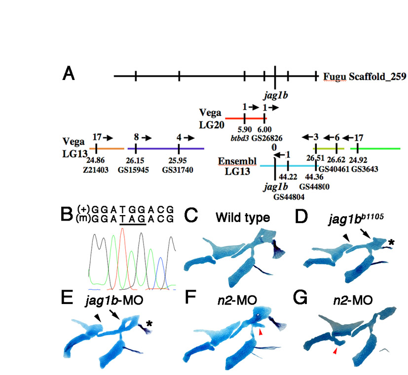
Fig. S1 Identification of the jag1bb1105 mutation. (A) Using synteny between Fugu and zebrafish Vega and Ensembl contigs, the b1105 mutation was mapped to a small region of LG13 containing jag1b. Recombinants per 2000 meioses are shown above each contig, and positions in Mb and marker names below. (B) An electrophoretogram showing sequence surrounding the G-to-A transition that creates a premature stop codon (underlined) in jag1bb1105 mutants (m). (C-G) Unilateral flat-mount dissections of 6 dpf facial skeletons stained for cartilage (blue) and bone (red). jag1b-MO larvae show similar skeletal defects to jag1bb1105 mutants, including truncation of Pq (arrowheads), shape changes in Hm (arrows) and transformed Op bone (asterisks). In notch2-MO larvae, ectopic cartilage processes (red arrowheads) are seen at low frequency near the DV interfaces of the hyoid (F) and mandibular (G) skeletons.
Image
Figure Caption
Acknowledgments
This image is the copyrighted work of the attributed author or publisher, and
ZFIN has permission only to display this image to its users.
Additional permissions should be obtained from the applicable author or publisher of the image.
Full text @ Development

