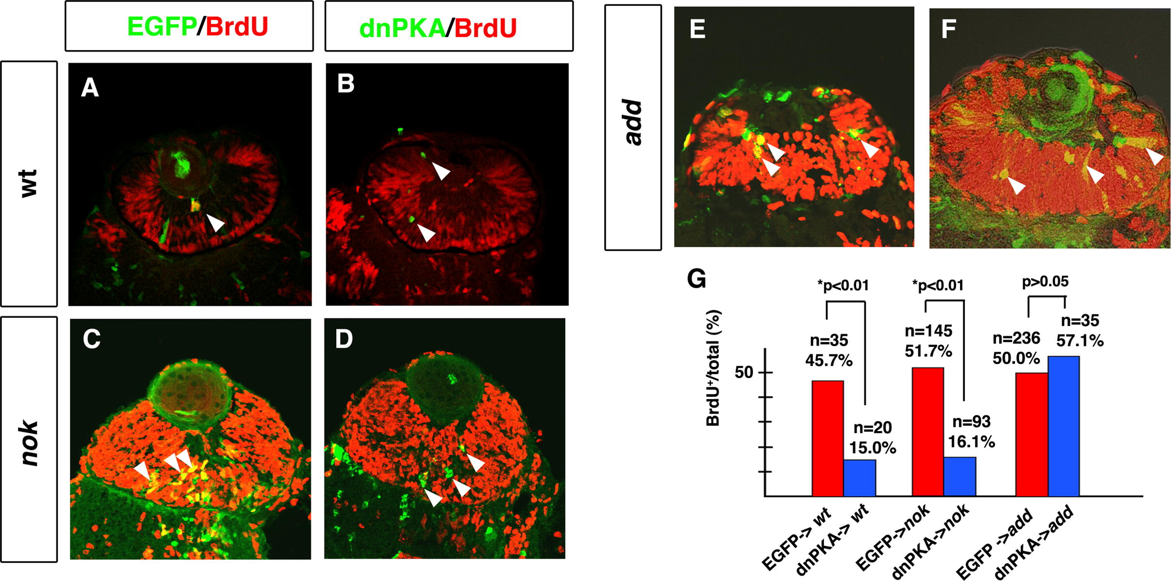
Fig. 7 dnPKA inhibits retinal cell proliferation in the nok mutant retina but not in the add mutant retina. (A and B) BrdU-labeled wild-type retinas expressing EGFP (A) and dnPKA (B). (C and D) BrdU-labeled nok mutant retinas expressing EGFP (C) and dnPKA (D). In both the wild type and nok mutant, retinal cells expressing dnPKA (green) do not incorporate BrdU (red) (B and D, arrowheads), whereas retinal cells expressing EGFP are BrdU-positive (yellow) (A and C, arrowheads). (E and F) BrdU-labeled add mutant retinas expressing EGFP (E) and dnPKA (F). Retinal cells expressing dnPKA (green) still incorporate BrdU (yellow) (F, arrowheads), similarly to retinal cells expressing EGFP (yellow) (E, arrowheads). (G) Percentage of BrdU-positive cells with respect to total number of retinal cells expressing dnPKA-GFP (blue bars) or EGFP (red bars) in wild-type retina and nok and add mutant embryos. The percentage of cells showing BrdU incorporation is significantly lower in retinal cells expressing dnPKA than in retinal cells expressing EGFP in wild-type retina and nok mutant retina (*p < 0.01: χ2-test). By contrast, the level of BrdU incorporation in add mutant retinal cells expressing dnPKA is similar to that in add mutant retinal cells expressing EGFP (p > 0.05: χ2-test).
Reprinted from Mechanisms of Development, 127(5-6), Yamaguchi, M., Imai, F., Tonou-Fujimori, N., and Masai, I., Mutations in N-cadherin and a Stardust homolog, Nagie oko, affect cell-cycle exit in zebrafish retina, 247-264, Copyright (2010) with permission from Elsevier. Full text @ Mech. Dev.

