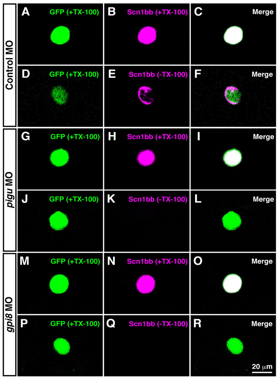
Fig. 6 Surface distribution of Scn1bb in RB neurons is diminished by knocking down GPI transamidase subunits. Dissociated RB neurons (GFP-positive) from 2 dpf embryos were labeled with anti-Scn1bb in the permeable (+TX-100) or impermeable (-TX-100) condition. (A-F) In control morpholino (MO)-injection, anti-Scn1bb labeled a whole RB cell in the presence of TX-100 (A-C), whereas it labeled the circumference of an RB cell in the absence of TX-100 (D-F). (G-I) Labeling with anti-Scn1bb with TX-100 displayed Scn1bb expression in an RB cell dissociated from a pigu morphant. (J-L) Scn1bb staining without TX-100 represented the impairment of Scn1bb surface distribution in a pigu morphant RB neuron. (M-O) Scn1bb labeling with TX-100 showed Scn1bb expression in a gpi8 morphant RB cell. (P-R) The surface expression of Scn1bb was not seen in an RB cell dissociated from a gpi8 morphant.
Image
Figure Caption
Figure Data
Acknowledgments
This image is the copyrighted work of the attributed author or publisher, and
ZFIN has permission only to display this image to its users.
Additional permissions should be obtained from the applicable author or publisher of the image.
Full text @ Development

